Rohini Pancholi-Bansal explores guided biofilm therapy (GBT), and asks if it’s a successful approach in the maintenance for a periodontal patient.
A 28-year-old transplant patient presented at Gipsy Lane Dental Practice, Leicester, with generalised periodontitis.
His clinical examination identified sites of oedematous swollen gingiva, fibrous surrounding tissue and marginal stubborn calculus (Figure 1).
His condition of a chronic advanced gum infection with generalised suppurating sites and extensive hyperplasia needed to be treated immediately for any possibility of stabilisation.
His predominant complaint upon arrival was bleeding gums and inability to access tooth surfaces during toothbrushing.
The patient was unaware of how severe his periodontal health had progressed. He showed determination to improve his current periodontal condition.
Following examination, we discussed what options there are to manage the condition. Opting for a course of guided biofilm therapy (GBT) was only considered once we had acclimatised the patient to what the treatment involves and explained its less invasive nature in comparison to non-surgical periodontal therapy (NSPT).
The patient also disclosed his anxiety levels with dental treatment and phobia from the surrounding dental surgery settings.
Prior to any clinical treatment. we spent a significant amount of time discussing what the various equipment is used for and how the patient is always in control of their treatment during its course. We used guided imagery to explain stages of treatment and focused on ‘tell, show, do’ and relaxation breathing exercises (Appukuttan, 2016).
The feeling of being overwhelmed and stress in the dental environment had previously led him avoiding dental care. Managing nervous patients requires strategic communication. Through various role play approaches and interactive methods, the patient became comfortable to continue and begin a course of GBT.
Biofilm removal
Recognising what GBT is and enforcing it for your patient and practice requires an in-depth knowledge on how this treatment differs from general NSPT.
GBT primarily focuses on the concept of dental biofilm removal. This is significant as dental biofilm is the main aetiological factor for caries, peri-implant and periodontal infections.
Biofilm is considered as a community or ‘layers’ of bacterial cells attached to a surface; in dental terminology, we identify it as plaque. The sticky, white yellowish ‘plaque’ that forms on the tooth surface and surrounding the gums is a type of dental biofilm (Marsh, 2006).
If not removed – due to inability to access specific sites, poor brushing techniques and failing methods – plaque can, over time, harden and form a substance known as calculus (tartar). Tartar can impede into the gingival margin and cause inflammation, swelling and exacerbate gum disease.
GBT was founded and created by Electro Medical Systems (EMS), a leading manufacturer in specific devices for dental prophylaxis. The original devices date back to 1983, but with modern technology and developments, EMS has been able to present a device that can be regarded as minimally invasive.
Airflow technology
To manage dental biofilm, debris and early calculus removal, guided biofilm therapy uses a specific prophylaxis device (called Airflow) combined with prophylaxis powder at a specific pressure and angle.
The Airflow technology cleans and polishes in a single procedure and can clean the gingival sulcus to a depth of 4mm.
There is also a Perioflow nozzle, which can be attached to the tip of the Airflow that will allow sub-gingival biofilm removal.
This nozzle is soft and streamlined for easy access into periodontal pockets between 4mm and 9mm. The flexible nature of the nozzle allows good adaptation into pockets, thereby increasing patient comfort.
The Airflow system is coupled with a compatible powder that took more than 30 years of research to create.
The use of the Airflow powder provides a perfectly smooth tooth surface where all the enamel prisms are kept intact. Polishing pastes can sometimes be abrasive to the tooth enamel, adding to any roughness on the surface. The high technology powders used for this treatment can vary from erythritol and sodium bicarbonate to glycine.
Treatment
The clinical practice of GBT focuses around an eight-step protocol cycle, which helps the patient to understand the treatment better, thus making it user-friendly.
Initially, we assess the patient, take a risk assessment and identify the severity of the patient’s periodontal health. We aim to give tailored advice specifically to each patient depending on their pattern of biofilm build up. This helps to engage better with the patient and improve bedside manner.
Generally, this initial stage provides a safety net for patients to feel engaged. At this stage my patient felt comfortable to ask questions regarding the treatment and what post-treatment advice entails.
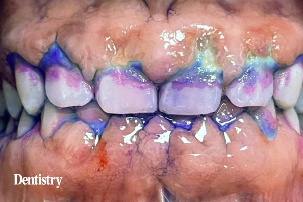
The next two stages work synergistically to motivate the patient with improving their oral health regime. We disclose the patient using plaque disclosing solution and a plaque score is taken. Together with the patient, we identify which areas they are missing when brushing.
Figure 2 illustrates plaque disclosed; the image shows a strong build-up marginally and interdentally, however the lingual sites were also abundant in plaque, the patient resulted in a plaque score of 72%.
At this stage, I discussed the relevance of reducing the plaque score below 40% and how this can be achieved through modified steps in their brushing routine.
Airflow in practice
The Airflow was then used – combined with the Airflow Plus Powder – to remove all the biofilm, staining and soft calculus, which was revealed at the disclosing stage. Following this, the Perioflow was inserted into sites of deep pocketing. However, due to the high level of inflammation and necrotic/decomposed gingival tissue build-up, it was difficult to fully access.
I used the Piezon Cavitron tip to remove the remaining stubborn calculus and treat any sites of burnished calculus. The Cavitron assisted in managing the necrotic gingiva, which was widespread on the buccal gingiva aspect of the lower mid sextant (Figure 3). This (pre- and post-) treatment image illustrates how in a single visit there is already better visibility of the tooth surfaces.
The use of a hand scaler and a Piezon is not essential or needed for each patient undergoing GBT. Each patient is specific and presents with differing challenges. In this instance, a Piezon was used generally around all four quadrants during treatment and a hand scaler, primarily a Gracey, was used anteriorly to manage the decomposed gum tissue and mature subgingival calculus.
I then carried out a final check to ensure the gingiva margins were free from any residual debris and sub-gingival calculus, a probe was used for detection. Finally, I sealed the dentition with a fluoride seal for enamel protection.
I asked the patient to return one-week post-treatment to assess for any sites of swelling, ulceration and infection.
We discussed a suitable recall time based on the patient’s risk assessment; a recall was scheduled for three months’ time to give the patient time to adapt to their OHR and for the healing phase within the gum tissue to stabilise.
Outcome
The patient attended one month after his treatment. He wanted me to assess if his gingiva tissues were healing as anticipated. One-month post-treatment, there is already successful signs in response to the treatment and healing (Figure 4a). Demonstrating both left and right sides of the buccal gingivae, there is reduced redness, improved exposure of the tooth surface, reformed gingiva contour and colouring.
With reduced redness and inflammation, the gum health is showing sites of marginal stippling and firmness within the tissues. The gingiva is showing positive results in healing due to the reduced bacterial load; we discussed there is good prognosis.
Pre- and one-month post-treatment (Figure 4b) indicates strong differences within the texture, composition and colour of the gingival tissue. The patient was content with his present results and motivated to reach remission.
I praised the patient’s efforts at this stage. I demonstrated a better angulation the patient can use to access his lower quadrant on brushing.
The patient is now due to return for his three-month recall appointment; I am hopeful the patient will present with a positive outcome.
What changes clinically?
Clinically, we carry over some elements of the traditional form of tooth cleaning . However, the long-term duration of the ultrasonic scaler, hand instrumentation with overlapping strokes and need for local anaesthetic are not essential with GBT.
With the option of GBT, we can provide and perform a treatment that does not feel intense nor traumatic for the patient yet is able to produce a better outcome.
With ever-growing adaptation to dental technology – such as with the use of the Perioflow to access pockets of up to 9mm to remove biofilm – and modernised equipment now available, patient comfort will not be compromised.
GBT positive factors
What benefits does GBT offer your patient? Feedback from GBT patients claim:
- Reduced sensitivity post-treatment
- Less pain during and after treatment
- Increased comfort during treatment
- Reduced trauma to the gum tissues
- Reduced recovering time after treatment
- Safer treatment in comparison to non-surgical periodontal therapy; due to reduced or no use of local anaesthetic
- Cleaner post-treatment feeling of the dentition
- No use of rubber cups, brushes or polishing paste, which can be confining and abrasive for the patient
- Reduced dental noises from hand-pieces in the surgery
- Reduced use of power and hand instrumentation.
Products used
- Guided biofilm therapy, Piezon Cavitron – EMS
- Gracey – LM Instruments
This article first appeared in Clinical Dentistry magazine. To read more like this, you can sign up to the magazine here.


