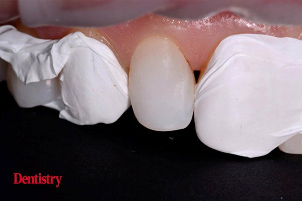
Linda Greenwall presents a case study utilising composite layering and direct filling following tooth whitening.
This case demonstrates the use of Ecosite Elements composite for a minimally invasive aesthetic smile makeover.
The 54-year-old female patient was keen to make improvements to her smile in a minimal invasive way. Several options were discussed to make smile improvements. These included tooth whitening, orthodontic treatment using braces or aligners and composite filling.
The risks, benefits, advantages and disadvantages of all techniques were discussed with the patient. It was decided to commence tooth whitening first, followed by composite filling.
The patient had previously whitened her teeth 10 years ago. She had old composite filling on both lateral teeth that needed to be replaced.
The existing composite restorations were stained around the margins and now needed to be upgraded.
There was a single tooth on the lower anterior teeth that had a slightly different colour to its neighbours. The patient explained that she had experienced minor trauma to this tooth 20 years previously and that it was asymptomatic.
An endodontic opinion was sought. This noted that this tooth was still vital and reacted to the electric pulp tester on the last reading.
It was decided that this tooth should be monitored. However, it could undergo tooth whitening treatment in the lower bleaching tray. The periapical radiograph showed that this tooth had pulp obliteration but no periapical lesion.
Whitening and planning
We undertook tooth whitening using 10% carbamide peroxide in a custom-made bleaching tray.
First, the upper teeth were whitened for two weeks. The patient was recalled for a shade review. It was noted that the upper teeth had reached a B1 shade after two weeks. Whitening was uneventful with no reports of sensitivity.
Lower whitening then commenced. Normally, a segmental bleaching tray can be used. However, it was decided that this was not necessary on this occasion. There was only a slight colour difference that could be whitened effectively.
Following whitening treatment (Figure 1), we waited for a period of two weeks to re-establish the bond strength of the enamel.
The patient wanted to ensure that her teeth looked natural and that her smile appeared as though she had not had anything ‘done’. Rather, she wanted an enhancement of her old composite filling.
She had considered porcelain veneers, but she did not want to have her healthy enamel cut down to prepare porcelain veneers. We undertook a simple diagnostic wax-up on the lateral and canine teeth. This showed the patient the new appearance of the teeth, which the patient approved (Figure 2).
A clear silicone index was made to the wax-up to help with the placement of composite material to replicate the shapes of the lateral incisors.
A silicone index (blue) was also made by the technician so that the patient could visualise the new appearance of the teeth (Figure 3). This was undertaken using Luxatemp temporary acrylic (DMG) bleached shade.
The Luxatemp was placed into the blue silicone index, which was then put on the patient’s teeth. After a setting time of two minutes, excess flash was removed.
The patient was pleased with the appearance of the mock-up teeth and gave her consent to proceed with the composite filling.
Composite shade choosing and layering
Once the Optragate (Ivoclar Vivodent) isolation is placed over the lips and the teeth are well isolated, composite shades can be selected (Figure 4).
This step needs to be undertaken rapidly as the teeth dehydrate quickly and so the incorrect shades can be selected: often, composite shades that are too light can be selected when the teeth dry out.
The material selected for this composite smile makeover was the Ecosite Elements from DMG.
This new composite offers flexibility to layer the teeth with different shades of enamel. This blends well with the shade of post bleached teeth and offers a high gloss finish.
Structure of the Ecosite composite
Ecosite Elements has pure set and layer set. The Ecosite Elements consist of (barium) dental glass in Bis-GMA based dental resins with filler content about 81wt%=65vol%. The filler size of the particles is 0.02-0.7um.
The Ecosite Elements has special properties in that silane is bonded into the glass surface and the other end, a double bond, is incorporated into the resin matrix helping for easy placement and light curing. This creates a chemical link between the filler and the resin.
Enamel that has been bleached changes in appearance; it can become more opaque and hence it is useful to use a variety of composite that has different properties of translucency and opacity for different parts of the tooth. Doing this can give a more natural appearance of the enamel after bleaching.
We then tested the shades of Ecosite directly onto the patient’s teeth.
The composite shade (Figure 5) was chosen immediately after the teeth were isolated, as once isolation commences the teeth dehydrate easily.
Composite shade selection
We selected two enamel shades – EB and EL – from the Ecosite Elements composite. There were several reasons for this:
The EB shade was selected as we wanted to enhance the bleached enamel and make it a feature of the mesiobuccal shape of this tooth to enhance this aspect
The EL lighter shade composite was layered over this for a natural, high-translucency effect
B1 was also selected as the true shade to bring everything together on the mesial section where the most composite needed to be added. There was a deficient mesial part and we needed to carry the true shade colour forward.
Layering technique
After cleaning the tooth prior to bond, etching was undertaken (Figures 6 and 7). PTFE tape was placed onto the adjacent teeth to protect them from any etch spillages.
Etch needs to be placed over the whole lateral tooth as the entire surface needs to be bonded in order to be brought, labially and mesially, into harmony for beautiful natural anatomy. The tooth was etched with 37% phosphoric acid (DMG Etch).
First, the B1 was used directly onto the mesial part of the tooth. Then EB followed as a secondary enamel lobe.
This was followed by EL shade, blended over to bring forward the glossy translucency of bleached enamel and to give a natural invisible transition from the natural tooth to the composite section.
Applying the silicone index
A transparent silicone index (Figure 8) was used in the case as it saves time, prevents air bubble formation and reduces composite defects.
The index assists with the correct shaping as this right lateral tooth needed to have a large mesial lobe section added onto the mesiobuccal part to match the contralateral tooth. This also saves time with polishing and contouring, as it avoids excess flash material being placed onto the tooth.
Firm pressure needs to be applied to the index once the composite is placed directly into it (Figure 9), which is why a test run is always helpful to check the layering shades and that the correct shade has been selected.
This approach is used often in the three-step technique when building up the occlusal vertical dimension of the bite in cases that have tooth wear from erosion, abrasion and attrition. It is used in aesthetic composite build-ups. This is because it can help speed up the building up the tooth in three dimensions.
Problems associated with poor placement in the index include excess material onto the adjacent tooth, overhang on the mesial side and a separation if not placed with Teflon tape or a clear matrix strip.
Final shaping contouring and finishing
Finishing touches were made to the composite restorations using a flame-shaped bur to ensure any excess composite was removed.
Once the secondary anatomy had been placed (Figure 11), the composite was polished using the ASAP (All Surface Access Polishers) polishing spirals. The interproximal flash was removed and smoothed using a finishing strip (Epitex) and floss was used to check there was no catching interproximally and on the mesiobuccal gingival area.
Finally, polishing of the composites was undertaken using the diamond-impregnated ASAP polishing spirals. These polishing systems come in two different colours and two sizes. A purple colour (44 microns) was followed by an orange colour (3-5 microns) for high shine final contour.
The spirals are useful as they allow for interproximal shaping without affecting the anatomy and finishing. This resulted in a smooth, glossy, glass-like surface that has a high shine (Figure 12).
This article first appeared in Clinical Dentistry magazine. To access more articles like this you can sign up to the magazine here.


