Home Dental Radiology Chronic maxillary sinus discomfort – The Journal of the American Dental Association

Chronic maxillary sinus discomfort – The Journal of the American Dental Association

by adminjay


To view the full text, please login as a subscribed user or purchase a subscription.
Click here to view the full text on ScienceDirect.

Figure 1

Panoramic radiographic exhibiting radiopaque/dense filling of the right maxillary sinus (arrow).

Figure 2

Maxillary sinus curettage revealed fragments of a mass of hyphae composed of an irregular mass of necrotic debris, bacteria, and filamentous organisms. (A) Hematoxylin-eosin stain (original magnification ×40). (B) Hematoxylin-eosin stain (original magnification ×400).

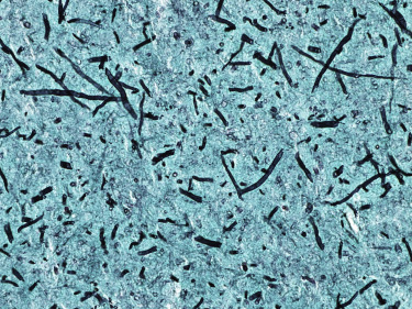
Figure 3

Gomori methanamine silver stain showed numerous filamentous organisms (stained black) resembling Aspergillus species. Fungal cultures confirmed the diagnosis of aspergillosis (original magnification ×400).

A 40-year-old woman had a 13-year history of right maxillary sinusitis. Her medical history was clinically significant for idiopathic, self-resolved chest pain and cesarean section. She described a history of extraction of an endodontically treated maxillary right first molar. She had been informed she likely had a remnant of the extracted tooth and possibly gutta-percha in her sinus, as suggested at the time by sinus opacification reportedly observed by radiographic imaging. Over time, she was treated with multiple courses of antibiotics, which provided only temporary relief of her sinusitis symptoms; she experienced recurrences of her symptoms shortly after completing each course.


Source link

Related Articles