TLD measurements
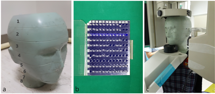
The head and neck components of a 5-year-old anthropomorphic phantom (ATOM® dosimetry phantom, model 705-D, CIRS, Norfolk, VA, USA), consisting of 8 slices with holes for TLD housing, were used (Fig. 1). The phantom was constructed to have equivalent tissue density to a human body measuring 110 cm in height and 19 kg in weight.
(a) Head and neck components of 5-year-old anthropomorphic phantom composed of 8 slices. (b) Thermoluminescent dosimetry (TLD) chips. (c) The phantom embedded with TLD chips was exposed to radiation using the paediatric mode of panoramic radiography.
TLD utilizes dosimeters that store radiation as energy and release it as light when stimulated by heat. The intensity of emitted light is converted into a value indicating the radiation dose via the reading unit. In this study, 48 pre-calibrated LiF TLD-700 chips (LiF 7: Mg, Ti) measuring 1/8 inches × 1/8 inches × 0.03 inches, with assigned identification (ID) codes, were used. Calibration was performed by a nationally certified company (ILJIN Radiation Engineering Co., Ltd, Gyunggi-Do, Korea) that maintains personal TLD badges through the following process. Dosimeters were exposed to 5612.7 µGy of radiation. Each dosimeter was read using a RADOS RE-1 reader (Rados Technology, Turku, Finland). The data were recorded and the sensitivity of each TLD detector was obtained. Since TLD was read as gamma energy which presents 1.25 times higher sensitivity compared to the x-ray, a correction factor of 0.8 was multiplied to normalize the value. TLD detectors with <±5% error were selectively used for this experiment.
Sixteen anatomic sites were selected and 3 TLD chips were placed at each site to minimize error. The dosimeter placement procedure was in accordance with previous studies6,11. The TLD chip ID and the anatomic site of each organ are summarized in Table 1.
The TLD-embedded phantom was exposed to panoramic radiography using an Orthopantomograph OP100 (Instrumentarium Imaging, Helsinki, Finland). Paediatric mode was selected, with exposure conditions of 66 kVp, 8.0 mA, and 16.8 seconds. The exposure conditions were chosen to correspond to the standard conditions used for 5-year-old children at Seoul National University Dental Hospital. Exposures were performed 3 times and averaged values were used to minimize error (Fig. 1).
The TLD chips were left for 24 hours, and the energy level stored in the chips was then read with a RADOS RE-1 reader (Rados Technology, Turku, Finland). Three unexposed chips were read to determine the amount of background radiation, which was subtracted from the results of the exposed chips. The absorbed dose of each anatomic site was obtained by averaging the measured value of the 3 chips in micrograys (µGy). The values from each anatomic site were integrated into the organ dose considering the tissue-irradiated fraction of the head and neck (Table 2). For example, the bone marrow dose was obtained by considering its distribution in the calvarium (11.6%), mandible (1.1%), and cervical spine (2.7%)12. Additionally, the bone surface dose was obtained by multiplying the bone marrow value by the bone-to-muscle attenuation ratio, which was defined as −0.0618 × kV(p) × 2/3 + 6.940613. The exposure of the skin, muscle, and lymph nodes was estimated to account for 5% of the total body tissue. The exposure fraction of the oesophagus was estimated as 10%. Other tissues of interest were counted as 100%. The individual organ doses were then integrated into the effective dose considering the tissue weighting factors suggested in 2007 by the International Commission of Radiological Protection (Table 2)14.
MC simulation
Monte Carlo (MC) simulation is an algorithm for predicting the interactions of X-ray photons with a complex medium, such as the human body15. When the appropriate physical and mechanical information is given, the organ-absorbed dose and effective dose can be calculated using computer software based on this algorithm.
Dose assessment in general conditions
For the MC simulations, PCXMC20Rotation (STUK, Helsinki, Finland), a supplemental program of PCXMC 2.0, was used. The virtual phantom of a 5-year-old in the program was 19 kg in weight and 109.1 cm in height.
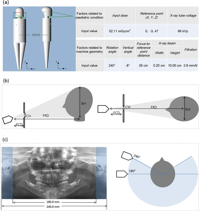
To obtain the absorbed dose and effective dose, the program required proper input values to be entered for the following factors: exposure dose, reference point, X-ray tube voltage, rotation angle, vertical angle of central ray, focus-to-reference distance (FRD), X-ray beam width/height, and filtration. The input values selected for the MC simulation corresponded to the same conditions as the TLD measurement method. The input values for the factors related to paediatric patients were determined as described below (Fig. 2a).

(a) The virtual 5-year-old phantom and the input values of the factors used to perform the Monte Carlo simulation. Schematic view of beam width, height (b), and rotation angle of the X-ray source (c) in panoramic radiography. Beam height and width were calculated based on the source-collimator distance, source-patient distance, collimator height, and collimator width. (FCD, focus-to-collimator distance; CH, collimator height; FRD, focus-to-reference distance; BH, beam height; CW, collimator width; BW, beam width). The rotation angle in panoramic radiography is correlated with the image length measured with a digital calliper. According to the manufacturer’s specifications, the rotation angle was 240°, which produced images of 240.0 mm in length. The possible minimum image length covering the lateral pole of the condyle was measured as 180.0 mm, which would correspond to a rotation angle of 180°.
Exposure dose
The dose-area product (DAP, mGy·cm2) is used for assessing the radiation dose of a diagnostic X-ray unit. The DAP of panoramic radiography in paediatric examination mode was measured using a DAP meter (Diamentor M4-KDK, PTW, Freiburg, Germany). The DAP meter was composed of an ionization chamber that was attached to the X-ray tube head and a set-top box displaying the DAP value. The measurement was performed 3 times and averaged to minimize error. The values were calibrated with temperature and pressure coefficients before being averaged.
Reference point
The reference point is the point where the central X-ray from all projection angles intersects, and it is shown in terms of X, Y, and Z coordinates. The X-axis crosses from left to right, the Y-axis from posterior to anterior, and the Z-axis from inferior to superior (Fig. 2a). The reference point was determined by the program to be (0, −3, 47), corresponding to the centre of the dental arch. The centre of the whole body was (0, 0, 0).
X-ray tube voltage
The X-ray tube voltage was 66 kVp, the same as the TLD measurement condition.
The input values for factors related to panoramic machine geometry followed the manufacturer’s specifications and previous studies in the literature that used the same machine (Fig. 2a)9.
Rotation angle
The rotation angle is the angle at which the X-ray source and the film rotate. The input value was 240°, according to the manufacturer’s specifications.
Vertical angle
The vertical angle is defined as the vertical angle formed by the central ray, and a value of −8° was entered.
FRD
A value of 35 cm was input for the distance from the X-ray source to the reference point.
Beam width and height
Beam width and height at the reference point were calculated based on the collimator size, FRD, and focus-to-collimator distance (FCD) (Fig. 2b)9. Values of 0.20 cm and 10.00 cm were input for the beam width and height, respectively. The equations for the calculation were as follows:
$$Beam,Height=frac{Collimator,Height,X,FRD}{FCD},$$
$$Beam,Width=frac{Collimator,Width,X,FRD}{FCD},$$
where collimator height = 3.78 cm, collimator width = 0.09 cm, FCD = 13.0 cm, and FRD = 35.0 cm. These values were obtained from the manufacturer’s specifications and manual measurements made using digital callipers.
Filtration
A value of 2.8 mmAl was input for filtration according to the manufacturer’s specifications.
Impact of individual factors on the effective dose
To assess the impact of each factor on the effective dose, the effective doses were repeatedly calculated by applying different values for each factor. When various values were input for a given factor, the other factors were fixed to the general conditions described above.
The minimum and maximum input values were determined for the rotation angle, vertical angle, FRD, X-ray beam width and height, filtration, and X-ray tube voltage based on the manufacturer’s specifications. Then, within this range, 6 input values were determined, at even intervals (Table 3). For factors that did not allow varied input values, including patient age, the reference point, and input dose, the standard values were used.
Rotation angle
The obtained panoramic image length measured with a digital calliper in the image viewer was 240.0 mm with 240° of rotation (Fig. 2c). The minimum projection angle was determined as 180°, from which a 180.0-mm length of image for a 5-year-old patient can be obtained (Fig. 2c). Rotation angles of 180°, 192°, 204°, 216°, 228°, and 240° were used.
Vertical angle
The vertical angle of the X-ray beam in panoramic radiography is known to range between −5° and −10°, so values of −5°, −6°, −7°, −8°, −9°, and −10° were input for this factor.
FRD
The patient’s position should not be closer to the source than the midpoint of the film-to-source distance (FSD) to obtain the image, considering the mechanical geometry of panoramic machines. Thus, the minimum FRD was determined as 25 cm, which was the half of the total FSD measured manually. The FRD was input with values of 25, 27, 29, 31, 33, and 35 cm.
Beam width and height
According to a report published by the Korean Ministry of Food and Drug Safety in 2014, collimator width was 0.2–4.5 mm and collimator height was 4.8–10.1 mm16. For the beam size calculation equation described above, the input values for beam width were 0.2, 1.06, 1.92, 2.78, 3.64, and 4.5 cm and those for beam height were 4.80, 5.84, 6.88, 7.92, 8.96, and 10.0 cm9.
Filtration
According to the 2014 report, in Korea, the mean filtration value was 2.66 ± 0.15 mmAl16. Thus, input values of 2.51, 2.57, 2.63, 2.69, 2.75, and 2.81 mmAl were used.
Tube voltage
The input values ranged from 57 to 72 kVp for this factor. The lowest available tube voltage for the machine was 57 kVp. To determine the maximum tube voltage, it was considered that male adults would be exposed to 73 kVp. Therefore, values of 57, 60, 63, 66, 69, and 72 kVp were input for this factor.
Data analysis
Statistical analysis was performed using IBM SPSS® Statistics version 23 (IBM Corp., Armonk, NY, USA). Simple linear regression analysis was applied to evaluate the influence of individual factors on the effective dose. The coefficient of determination (R2) was obtained to verify the fit of this analysis. Regression coefficients were obtained to compare the impact of each factor on the effective dose.


