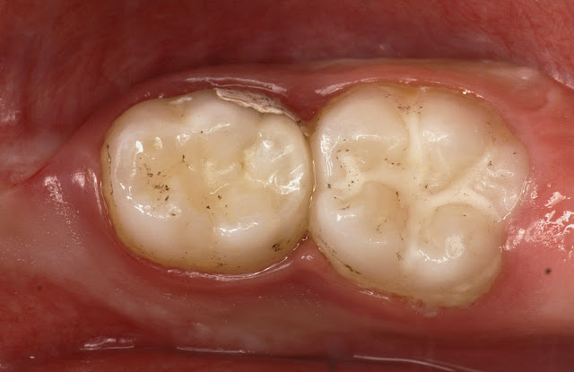
Use of Glass Ionomer Cements in Paediatric Dentistry: Clinical Cases of Application in Primary Teeth
Introduction
Successful restoration is linked to various factors: the material, the practitioner and the patient (Donovan et al. 2006). The latter characterises the uniqueness of paediatric odontology. The patient’s (sometimes limited) co-operation justifies the use of materials that can be easily manipulated and that are favourable to a simple protocol. Furthermore, primary teeth are distinguished from permanent teeth mainly by their anatomy and their limited time in the dental arch.
Consequently, even if the practitioner has the same array of materials for permanent teeth as for primary teeth (composite resins, amalgams, compomers and glass ionomer cements (GICs)), the specificities for the restoration of primary teeth are different.
After reviewing the uniqueness of primary dentition, a summary of current data in the literature on the longevity of GICs in this clinical indication will then be presented, followed by a discussion of GICs modified by the addition of resin (RMGICs) and packable GICs (pGICs). Finally, the principal uses of these cements will be illustrated by examples of clinical cases. Composites modified by the addition of polyacids (or compomers) will not be discussed in this article because these are more similar to composites than glass ionomers.
Criteria for Selecting a Material in Paediatric Odontology
This section is limited to criteria concerning the characteristics of primary teeth and the types of caries.
Primary teeth are characterised by a thin layer of enamel consisting of enamel prisms that are directed vertically to the proximal surface. In the case of carious lesions, this tenuity can lead to extensive destruction, exacerbated by the fact that the prisms have poor cohesion. Dentin forms an equally thin layer and its wide tubules allow bacterial penetration, accelerating the risk of pulp contamination. It is therefore important to work with sealable restorative materials.
The pulp chamber is proportionally much bigger than in permanent teeth and the pulpal horns are prominent. A carious lesion can therefore occur rapidly close to the pulp. It is therefore important to work with adhesive materials that do not require secondary cavity retention forms that may decay and cause pulpal exposure. For the same reason, smooth surfaces in the youngest patients which are affected by linear enamel caries or early carious lesions in the occlusal grooves or proximal surfaces of molar teeth (Psoter et al. 2003; Psoter et al. 2009) call for minimally invasive adhesive dentistry.
Owing to their short crown height, marked cervical constriction, relations with adjacent teeth and large gingival papillae, primary teeth can cause difficulties in establishing an isolated operative field, rendering the use of hydrophobic materials problematic (Burgess et al. 2002). It is therefore important to work with hydrophilic materia
Proximal caries adjacent to the primary tooth under treatment are common. Fluoride-releasing material placed on the proximal surface of the restoration could be advantageous in a favourable environment, in patients with a controlled risk of caries, to reduce the development and progression of caries on the proximal surface of the adjacent tooth. It is therefore important to work with bioactive material (Qvist et al. 2010).
Moreover, the tooth’s sometimes short remaining time in the arch may admit the use of materials compatible with this duration. Additionally, as masticatory constraints in children are lower than in adults (Braun et al. 1996; Castelo et al. 2010; Palinkas et al. 2010), materials that are relatively less mechanically resistant may prove to be suitable. Thus, while materials with mechanical properties are crucial for permanent teeth, materials with lower mechanical properties may suffice for primary teeth in certain situations. This explains why glass ionomers, markedly less mechanically resistant than composites, may have a role in paedodontics.
Therefore, besides the need for fast implementation related to the patient’s age, restorative material for primary teeth should also be sealable and adhesive to tooth tissues, bioactive and hydrophilic.
Glass ionomers meet all of these requirements.
Longevity of Restorative Materials in Primary Teeth
A review of the literature concerning the longevity of dental materials used in primary dentition highlights a wide variation in success rates. Indeed, numerous factors are involved: the type and brand of the material used, the practitioner’s experience, the site and the depth of the carious lesion, as well as the age and co-operation of the patient.
Additionally, the life span of restorations in primary teeth is significantly different from that of permanent teeth, regardless of the chosen material (Hickel and Manhart 1999). This emphasises the specificity of the selection criteria for primary dentition material.
Yengopal and coll. in 2009 (Yengopal et al., 2009) conducted a systematic review of the literature, comparing the outcomes of different materials used for the restoration of primary teeth, in terms of pain relief, durability and aesthetics. The study concluded that, from 1996 to 2009, there were only two well-conducted randomised clinical trials evaluating the different restorative materials. These trials reported no significant differences between the materials.
In one of these two trials, Donly and coll. in 1999 (Donly et al., 1999) compared a RMGIC (Vitremer®) with amalgam over a three-year period. However, owing to the high “lost to follow-up” rate, only the 12-month results are reported. No significant difference was found.
In terms of longevity, GICs are therefore materials that may pose an alternative to amalgams or composites for the restoration of primary teeth for a limited period of time.
At present, two types of GIC are clinically relevant: RMGIC and pGIC. However, some studies demonstrate differences in longevity depending on the type of GIC used and the site (occlusal or proximal) of the cavity.
The Two Main GIC Types
Of the different GIC types, two are particularly suitable for paediatric dentistry:
1) RMGICs (resin-modified glass ionomer cements)
Fuji II® LC (GC), Riva Light Cure (SDI), Photac-Fil® (3M-Espe), Ionolux (Voco).
2) pGICs (packable glass ionomer cements)
Fuji IX (GC), Riva Self Cure (SDI), HiFi (Shofu), Ketac Molar (3M-ESPE), Chemfil Rock (Dentsply) or Ionofil Molar (Voco).
The main differences between these two types of material relate to their mechanical properties and implementation.
RMGICs demonstrate moderate resistance to wear, but this is sufficient for restorations that have a fixed time in the dental arch. Qvist and coll. (Qvist et al., 2010) report that the longevity of RMGICs is almost equal to that of amalgams, but is higher than that of pGICs. These materials may be indicated for occlusal and proximal restorations in primary teeth, for a remaining period in the arch of around three to four years (Qvist et al. 2004; Courson et al. 2009). The implementation of RMGICs is often favoured by practitioners, given the possibility of curing by photopolymerisation.
Packable GICs have the advantage of single-step placement (particularly attractive property for proximal cavities) and, in certain formulations, have accelerated chemical bonding. However, they are not robust in the medium term in proximal areas (Qvist et al. 2010). Limiting their use proximally for less than two to three years in the dental arch, and also for their use in small to medium sized cavities, is therefore recommended (Forss and Widstrom 2003). They can also be used for larger multi-sided cavities, but in this case covered with a pre-shaped paedodontic crown (Courson et al. 2009).
Nevertheless, it is possible that the use of a protective varnish (G-Coat Plus®, GC) may considerably improve durability as shown by a recent study by Friedl et al. in 2011 (Friedl et al. 2011), which concluded that these materials could be used for permanent posterior restorations. However, one might question how bioactive fluoride-releasing properties are maintained when a protective varnish is used.
Finally, it should be noted that a new high-viscosity RMGIC is now available (HV Riva Light Cure – SDI); this is a RMGIC that can be used as a pGIC.
Examples of Clinical Cases
Whatever the clinical situation, an operative field will always be established whenever possible. For the following two clinical cases, in which the lesions are not easily accessible, an isolated operative field was established. It should be noted that, with or without an operative field, the bioactive nature of GICs, with their fluoride release, gives them an advantage over adhesive materials.
Clinical case no. 1 (Dr. L Goupy)
Example of the restoration of a proximal and cervical lesion on a primary tooth with a RMGIC:
Fuji II ® LC (GC).


In this case involving a juxta-gingival buccal lesion, RMGIC was the appropriate procedure. Admittedly, a composite restoration could have been carried out proximally as the operative field could be established. However, for practical reasons, we decided to use the same material so as to avoid having two different protocols to restore the same tooth.
Clinical case no. 2 (Dr. L Goupy)
Example of occlusal restoration on a primary tooth with a packable GIC: Riva® Self Cure (SDI)


This second case is entirely different from the first. It involved a very young child with early childhood caries. The use of GIC material is indicated in this case in point, as the bioactive properties of the material are especially useful.
Conclusion
The principal characteristics of glass ionomers include the ability to adhere naturally to enamel and dentin, the cariostatic effect of fluoride release and moisture tolerance. They are therefore particularly worthwhile materials for use in challenging clinical situations concerning unco-operative children or even when isolation is impossible to obtain due to the anatomical peculiarities of the primary teeth. In this regard, either a RMGIC or a pGIC would be used when mechanical stress, particularly due to wear, will be great. In a future article, we shall discuss the role of compomers in paediatric dentistry in relation to these materials.
References
– Braun S, Hnat WP, Freudenthaler JW, Marcotte MR, Hönigle K, Johnson BE. A study of maximum bite force during growth and development. Angle Orthod 1996; 66: 261–4.
– Burgess JO, Walker R, Davidson JM. Posterior resin-based composite: review of the literature. Pediatr Dent 2002; 24: 465-79.
– Castelo PM, Pereira LJ, Bonjardim LR, Gavião MB. Changes in bite force, masticatory muscle thickness, and facial morphology between primary and mixed dentition in preschool children with normal occlusion. Ann Anat 2010; 192: 23-6.
– Courson F, Joseph C, Servant M, Blanc H, Muller-Bolla M. Restauration des Dents Temporaires [Restoration of primary teeth]. Encycl Med Chir, Odontologie 2009; 23-410-K-10.
– Donovan TE. Longevity of the tooth/restoration complex: a review. J Calif Dent Assoc 2006; 34: 122-8.
– Donly KJ, Segura A, Kanellis M, Erickson RL. Clinical performance and caries inhibition of resin-modified glass ionomer cement and amalgam restorations. J Am Dent Assoc 1999; 130: 1459-66.
– Forss H, Widström E. The post-amalgam era: a selection of materials and their longevity in the primary and young permanent dentitions. Int J Paediatr Dent 2003; 13: 158-64.
– Friedl K, Hiller KA, Friedl KH. Clinical performance of a new glass ionomer based restoration system: A retrospective cohort study. Dent Mater 2011; [Epub ahead of print].
– Hickel R, Manhart J. Advances in glass ionomer cements. Glass-ionomers and compomers in pediatric Dentistry. Berlin, Quintessence, 1999.
– Palinkas M, Nassar MS, Cecílio FA, Siéssere S, Semprini M, Machado-de-Sousa JP, Hallak JE, Regalo SC. Age and gender influence on maximal bite force and masticatory muscles thickness. Arch Oral Biol 2010; 55: 797-802.
– Psoter WJ, Zhang H, Pendrys DG, Morse DE, Mayne ST. Classification of dental caries patterns in the primary dentition: a multidimensional scaling analysis. Community Dent Oral Epidemiol 2003; 31: 231-8.
– Psoter WJ, Pendrys DG, Morse DE, Zhang HP, Mayne ST. Caries patterns in the primary dentition: cluster analysis of a sample of 5169 Arizona children 5-59 months of age. Int J Oral Sci 2009; 1: 189-95.
– Qvist V, Poulsen A, Teglers PT, Mjör IA. Fluorides leaching from restorative materials and the effect on adjacent teeth. Int Dent J 2010; 60: 156-60.
– Yengopal V, Harneker SY, Patel N, Siegfried N. Dental fillings for the treatment of caries in the primary dentition. Cochrane Database Syst Rev 2009; 2: CD004483.
by Dr. Elisabeth Dursun, Dr. Lucile Goupy, Dr. Frederic Courson, Dr. Jean Pierre Attal


