Home Dental Radiology Intraoral and extraoral hyperpigmentation and an expansile mandibular lesion

Intraoral and extraoral hyperpigmentation and an expansile mandibular lesion

by adminjay


To view the full text, please login as a subscribed user or purchase a subscription.
Click here to view the full text on ScienceDirect.

Figure 1

Radiographs of long bones. Selected plain radiographs of the distal humerus (A) and proximal femur (B) exhibit a change in the quality of bone, mimicking ground glass.

Figure 2

Histologic presentation of tissue submitted after recontouring of expansile mandibular lesion. A. A low-power (hematoxylin-eosin, original magnification ×40) image of the tissue specimen shows irregular trabeculae of woven bone in a somewhat parallel arrangement within a fibrous connective tissue stroma. There is a marked clefting artifact separating the connective tissue stroma from the bony trabeculae. B. A higher power (hematoxylin-eosin, original magnification ×100) view of the tissue shows the minimal osteoblastic rimming around the trabeculae in scattered areas but absent entirely in others. The fibrous tissue stroma, parallel trabeculae arrangement, and cleft artifact can also be seen at this power.

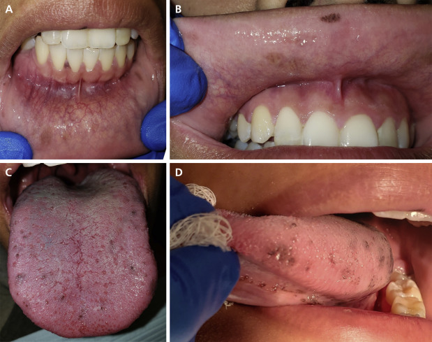
Figure 3

Clinical presentation of pigmented macules. A. Multifocal pigmented macules of varying degrees of pigmentation noted on the lower labial mucosa and on the gingival papillae between teeth nos. 25 and 26. The buccal expansion of the right mandibular alveolar ridge can be seen as well. B. Pigmented macules of the upper labial mucosa include a smaller, dark macule near the midline and a faint, subtle, larger macule on the mucosa adjacent to tooth no. 7. C. The dorsal tongue exhibits multiple hyperpigmented papillae. The subtle blue coloration is due to the staining from chewing gum that the patient was chewing before the examination. D. Pigmented macules are noted on the dorsolateral and ventrolateral tongue.

Figure 4

Clinical presentation of mandibular buccal and lingual expansion.

Figure 5

Panoramic radiograph showing mandibular expansion and bony changes. An expansile lesion of the right mandible that has caused tipping of the roots of teeth nos. 27 and 28 is evident. This lesion exhibits a ground-glass quality and extends from approximately tooth no. 20 through tooth no. 30. The radiographic change is more subtle in the maxilla.

A 19-year-old woman was referred to the Oral and Maxillofacial Pathology Resident Clinic at Northwell Health Long Island Jewish Medical Center with a primary symptom of “dark spots on my tongue and lips.” She initially reported a noncontributory medical and social history and was not taking any medication at that time nor had any allergies to report. On further questioning, the patient revealed a prior diagnosis of a condition involving multiple bones, including her femur, humerus, maxilla, and mandible.


Source link

Related Articles