

With tighter COVID restrictions on the horizon, Emma Laing explores how she will keep her orthodontic patients ticking over.
Where we are now?
I write this after watching the news strongly indicating that a second lockdown is coming to England in the near future.
Whether or not this happens I feel there is certainly an air of ensuring treatment efficiencies are made with our dental treatments. For example, booking appointments as soon as possible and ensuring we do the most at each appointment for each patient.
I am consciously scanning the schedule and fallow times to see if there are any spaces to fit patients in.
Sadly I suspect the winter may cause restrictions in the way we work.
Orthognathic cases
The surgeon I work with has limited operating slots when compared with the pre-COVID period.
The hospital she uses was, like many others, reallocated for NHS use during the pandemic. It has not yet resumed a full private schedule.
So we’ve been closely collaborating planning cases. For now we are setting October dates for our patients waiting to go ahead.
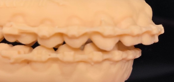
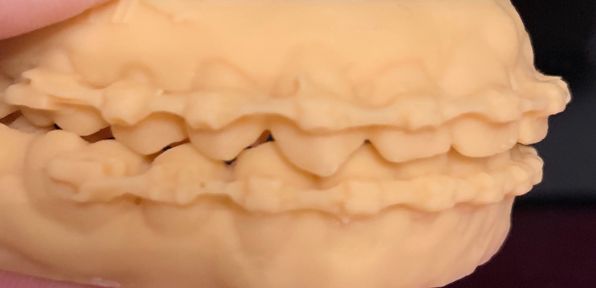
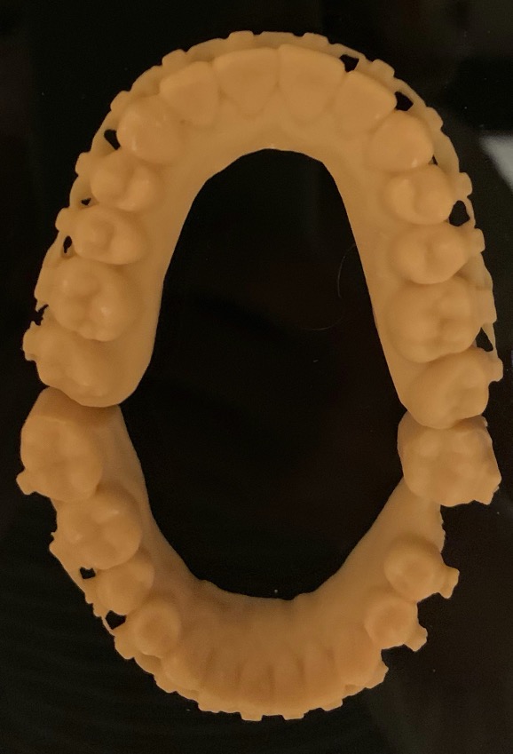
As previously written I love my new Itero Flextech scanner. It is compact and super accurate. The image quality gives a wow factor for patients and gives me additional information for each case.
For orthognathic cases, I have recently been using it to scan the arches with fixed appliances in situ. This way I can have digital study models 3D printed prior to orthognathic surgery (Figures 1-3).
The accuracy is incredible; you can even see the Damon bracket morphology and the tiny hooks.
Plaster models taken in this situation were often covered with remnants of red modelling wax where the brackets were blocked out. This often led to distortions in the models and the surgical wafers produced.
The surgeon and I are delighted with the printed models, which are both accurate and reproducible.
We can forward the files to the technician making the wafers in a different lab with ease.
Dental photography
I was so happy to attend a dental event in person last week – Dr Minesh Patel’s excellent F:ocus course.
Well organised with social distancing and mask wear, it was a pleasure to be in the company of dental colleagues and hone our skills after months of none of the usual dental meetings and conferences.
Minesh discussed various skills and techniques. Specifically covering the use of photography in communicating with labs. He highlighted the role of lighting sources in this.
I thought my photography was quite good until I saw his results. I left completely motivated and armed with a shopping list of kit to invest in.
Using the excellent MPB website, I have now therefore changed my 100mm macro lens that I know most orthodontists use, to a 60mm one.
Unbeknown to me it was too large for the cropped sensor of my 700D Canon body.
I am using diffusers for intra-oral photographs and a ‘portrait kit’ for extra-oral photographs.
These additions elevate my photography. And they have also been a great talking point with patients.
I’ve now been passed on a lot of safari photography ideas with my kit from one patient, which sound amazing if we are able to travel in 2021.
Going digital
For now I am honing in on filling my inbox every day with digital dentistry. The advancements are great to hear.
From Invisalign bringing out the Virtual Care concept for monitoring case progress, through to the advances in the detailed Dental Monitoring system.
At least we can use these tools during the inevitable restrictions to come.
Follow Dentistry.co.uk on Instagram to keep up with all the latest dental news and trends.