This study utilized a wide dataset and employed a data-driven analysis process to produce objective results. The findings on normal occlusion in Koreans hold significant value for future studies on malocclusion in this ethnic group, as oral and maxillofacial characteristics and malocclusion patterns can vary among different ethnicities41. In addition, the results revealed several significant issues.
Firstly, this study’s analyses cover a more extensive range than previous studies, but they exhibit consistency in overlapping areas. Secondly, interpreting the geometric aspects of variables enables us to distinguish subtle differences between variables that are known to represent the same feature in clinical practice. Thirdly, there were instances where variables known to represent different traits were linked eventually to the same anatomical feature. Finally, through the analysis of the correlation structure of the cephalometric measurement set, we identified nine key characteristics of the oral and maxillofacial region. We also identified clusters of variables that exhibit these characteristics and investigated the inter-correlations between the groups.
In order to confirm the validity of the findings in this study, a comparison with previous studies revealed a significant level of consistency. Most previous researches have focused primarily on class III malocclusions in young patients, which makes it challenging to compare with adult malocclusions analyzed in this study. The only exception was a study by Scala et al. that included a section on adult females with Class III malocclusions28, and we were able to compare their results with ours. Out of the 17 variables that were measured in the prior study, 14 either were included or have a significant similarity to the 65 variables examined in this study. Therefore, we constructed an unweighted network (with a cutoff at 0.6) to assess the 91 correlations between those 14 variables. As a result, it was confirmed that the structure was completely identical, except for one case (Wits—ANB). These findings are deemed acceptable because the variables associated with maxillofacial anteroposterior discrepancy may exhibit different patterns within the class III malocclusion and normal occlusion groups. So, there is a high degree of consistency between the two studies. This comparison covers only 4.38% of the variable pairs examined in this study, but this is because the current study is based on a broader data set than previous studies.
ANB and Wits appraisal, previously mentioned as exceptions, are commonly used as clinical indicators representing the degree of maxillomandibular anterior–posterior positional discrepancy. A comparison was made between the correlation coefficients of four indices: [17] A-B plane angle, [23] ANB, [31] Wits appraisal, and [62] APDI ([No.] refers to the corresponding cephalometric variable in Table 1.). All four were clinically accepted as representative of the same anatomical feature. It was found that the first two variables had a strong correlation of 0.95. However, Wits-ANB, which also falls into this category, had a relatively lower correlation of 0.56. To further investigate these aspects, the correlation structure of these four variables and other closely related variables with strong correlations was analyzed in detail.
Despite the fact that the four variables are clinically known to represent the same anatomical feature, two of them exhibit a strong associate structure with the facial convexity, one has moderate correlations, and one does not belong to the cluster. Although the four nodes mentioned earlier exhibit correlated structure in the lower left of Fig. 3, they do not form a single cluster. Group C, represented by the red square nodes and including two of the four nodes, is a cluster exhibiting strong correlation (> 0.8) between two maxillofacial anterior–posterior disparity variables ([23] ANB and [17] A–B plane angle) and two anterior facial convexity variables ([4] convexity of point A and [16] facial convexity). In this case, a group of nodes in which each node is strongly connected to every other node is known as a clique structure28. In addition, Fig. 2a shows that [31] Wits appraisal and [62] APDI, which are not included in Group C, show differences in their connectivity. APDI is not strongly connected to group C, but forms a clique structure with moderate correlations (0.63–0.69) with all nodes in this group. On the other hand, [31] Wits appraisal showed weak correlations (0.47, 0.47, 0.56 and 0.62) with the corresponding group, leading to its exclusion from the cluster.
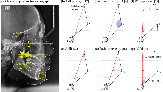
As can be seen in Fig. 4a, we considers point B and the pogonion to be in approximately the same position from an anterior–posterior perspective, and a geometric analysis was conducted for the six indices. In all the cases of Fig. 4b–e, the anteroposterior position of the point A is evaluated in relation to the vertical plane of the anterior surface. However, it can be divided into three cases according to the reference planes. Figure 4b–e shows that [17] A–B plane angle, [23] ANB, [4] Convexity of point A and [16] Facial convexity all use the facial plane (N-pog.) as the vertical reference line. Furthermore, a geometric relationship of the [31] Wits appraisal depicted in Fig. 4f, shows that it estimates the anteroposterior position of the point A relative to the perpendicular of the occlusal plane passing through point B. On the other hand, the [62] APDI index is defined as the sum of the [11] Facial angle, [17] A–B plane angle, and [14] palatal plane angle21. Alternatively, the angle of the A–B plane in relation to the palatal plane can replace it based on the geometric relationship42. Next, this index is converted into a measure of the anteroposterior position of point A to a line perpendicular to the palatal plane passing through point B, as shown in Fig. 4g.

Geometric analysis of indices evaluating the anterior–posterior positional relationship of the maxilla and mandible and neighboring variables on the network. (a) Major landmarks & reference planes on a lateral cephalometric radiograph, (b) A–B plane angle, (c) ANB, (d) Convexity of point A, (e) Facial convexity, (f) Wits appraisal, and (g) 90° – APDI.
By geometrically transforming and interpreting the variables representing the degree of convexity of the anterior face and the variables representing the anteroposterior positional relationship of the maxilla and mandible, it was found that variables known to represent the same anatomical characteristics in clinical practice are sometimes subdivided and variables representing different characteristics are sometimes grouped together. The difference between the correlation configurations of the [31] wits appraisal and the [62] APDI is also interpreted in this way. The landmarks, N and the palatal plane, which determine the vertical reference planes, are equally located on the skeletal structure, so that the [62] APDI shows a moderate correlation with the variables in group C. However, the occlusal plane, which is the reference of the [31] wits appraisal, is a dental structure, so that it is estimated that it shows a low correlation.
The inter-correlation structure among clusters applying cutoff value of 0.6 in Fig. 2a supports the presence of nine major anatomical characteristics in cephalometric variables. All groups, except for group A-1, exhibit a weak inter-correlation with each other. This indicates that the primary anatomical features represented by groups A-2, B, C, E, F, G, H, and I, whose meanings are described in Table 2, are independent features. The anteroposterior positions of the maxillary and mandibular incisors in relation to the facial anterior part (Group A-1) displayed significant density correlation with Group A-2 and high relative connectivity with Groups B and C, likely due to their proximity to anatomical structures. In classical cephalometric analysis, the variables of A-1 are viewed differently, often used as auxiliary measurement variables or considered independently17,18,19, while some studies pair them with groups (A-2, B) that measure the anterior angle7,8,11,12,13. Although A-1 is highly correlated with adjacent groups, it can still be considered an independent anatomical characteristic as there is a practical advantage to studying it separately, based on the network structure.
By applying the MST algorithm to the network structure illustrated in Fig. 3, we can directly grasp the key pairs of variables in the connections between the 10 highly clustered groups of variables, and more information can be obtained by considering the information in Fig. 2a together. The intra-correlations of each cluster exhibit a strong clique or clique-like structure, except for Group D. That is, each of the nine dense clusters independently represents an anatomical feature in the oral and maxillofacial region, which is the main characteristics revealed by various cephalometric analyses.
Group D should not be considered a single anatomical feature because, unlike other groups, it does not have a clique or a cluster structure similar to a clique. Kim’s study introduced the [60] Overbite depth indicator (ODI) which consists of a combination of two variables14,16,21, [61] A–B to Mandibular Plane and [14] Palatal Plane angle, and it represents a single anatomical characteristic, the depth of the overbite. However, in the case of [63] combination factor or [64] Extraction index, it is designed as an index that mixes different anatomical characteristics to be used as a basis for decision-making. In Group D, each variable is a subset of the other by definition. Therefore, the strong correlation is limited to the edges of one line and cannot indicate the same anatomical feature. In summary, the analysis of 65 cephalometric measurements showed that nine anatomical characteristics were the primary factors, except for group D as presented in Table 2.
Statistical and advanced network analysis methodologies, weighted network and minimum spanning tree, were employed to visualize the correlation structure among anatomical characteristics in this study. This study process can be standardized and applied as a framework for studying malocclusions of Class I, II, and III as well as different ethnicities. Comparing the outcomes of studies that use identical data sets and analysis framework is expected to improve the value of future research.

