The aim of this meta-analysis was to determine whether the craniofacial cephalometric characteristics of individuals with DS differ from those of the general population.
It was decided not to limit the literature search by language or date of publication in order not to miss any article that might provide information relevant to the work’s hypothesis; as a result, articles in three languages were included.
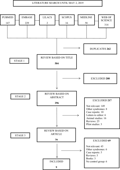
When evaluating the comparability of the cases and controls by means of the NOS, the age and ethnicity of subjects were considered, as cephalometric measurements may be influenced by these variables. Although in the seven works included for meta-analysis, age ranges and ethnicity were the same in case groups and control groups, there was some variability between the articles, especially regarding age, so in order to maintain homogeneity, the works were grouped according to age ranges into two groups: 6–11 years27,28 and 5–22 years22,23,24,25,26.
The seven studies included in meta-analysis used different cephalometric measurements, and so it was decided to analyze only those measurements that were repeated in at least three articles. Nevertheless, the maximum number of works to coincide in any single measurement was five. For this reason, the results of the present meta-analysis should be interpreted with caution.
Cranial base length was evaluated by means of the measurements SN, BaS and BaN. It was found that anterior cranial base length (SN) was significantly shorter in DS subjects than among control subjects. Some authors have suggested that the lesser development of the anterior cranial base is related with smaller brain size27. But according to Enlow the growth of the cranial base is considered rather autonomous, as compared to the cranial vault, because of that, we think that the smaller brain size of DS individuals should not be considered the main cause or the only cause of the short anterior cranial base of these patients. Rather both facts could be the effect of a different primary cause30. One study evaluated cranial base growth in subjects aged 8–18 years, observing that it grew in the same proportion among DS individuals as control subjects, showing that the deficit in size was produced before the age of 8 years22. Some authors have suggested that the deficit is of prenatal origin9, while others state that is develops during the first 4 years of life31.
As for posterior cranial base (BaS), it was also found to be significantly reduced in size in DS groups. A histological study based on autopsies of individuals at different ages determined that growth in this region comes to an end at the age of 18 years in healthy individuals32. Alió et al. observed that, in individuals with DS, the rate of growth decreases gradually up to the age of 15 years, while in control subjects it continues to grow, so that spheno-occipital synchondrosis growth stops earlier in individuals with DS22.
Obviously, the reduced anterior and posterior cranial bases in cases of DS mean that the total cranial base length (BaN) is also significantly smaller than in the general population.
At the same time, the cranial base angle (SNBa) was significantly larger in DS than control subjects. It has been suggested that delay in intra-sphenoidal synchondrosis fusion in postnatal life is essential to the cranial base’s flattening mechanism. Radiographic data obtained from individuals with DS show a delay in fusion, so that synchondrosis remains without fusing between the ages of 1–7 years, while in the general population it is fully obliterated by the end of the first year of life33. The present findings show that the difference in measurement between DS cases and control subjects was more acute in the 5–22 year age range. But the difference in effect size between the 6–11 year and the 5–22 year age group was 2.35°, so even though this difference was statistically significant, it was not clinically significant, as the standard deviation for this angle is around 4°34. In addition, according to Bjork, the cranial base angle is 130.8° ± 4.2° at age 12 and 131.6° ± 4.5° at age 20 years, showing an insignificant change from the former age to the latter34. Almeida et al., in their systematic review showed that SNBa remains constant from 5 to 15 years of age35. Alió et al. observed that in both individuals with DS and the general population it does not vary between the ages of 8–18 years22.
The anteroposterior position of the maxilla was evaluated by means of the SNA angle, which, although smaller in cases of DS, did not show significant differences in comparison with control subjects. The SNA angle relates the maxilla to the cranial base, which, as stated above, is significantly shorter in individuals with DS. Anteroposterior and vertical cephalometric measurements for the maxilla and mandible based on an anomalous cranial base can lead to erroneous cephalometric interpretation. For this reason, Jesuino and Valladares suggest that in these cases cephalometric measurements should take the Frankfort plane as reference27. The reduced anterior cranial base in DS can make the SNA similar to that of control subjects due to a geometric effect, even though the maxilla is smaller23, because the N point will be located at a more posterior location24.
Maxillary length (CoA) was significantly smaller in DS than control groups, corroborating the existence of maxillary hypoplasia in the sagittal plane. Alió et al. showed that the maxilla grows in the same proportion between the ages of 8 and 18 years in DS cases as in healthy subjects24. Klingel et al. observed that at the age of 6–9 months the maxilla is already smaller in all three dimensions in DS infants36.
As for the mandible, no statistically significant differences were observed between DS and control subjects in SNB angle, although values were higher in DS subjects. This measurement is also related to the cranial base; in this case shortening produces larger SNB angles23. At the same time, the present work found that the cranial base angle was significantly larger in DS groups, which would lead to less symphysis projection, while in control subjects the angle was more acute, contributing to a more anterior mandibular position35. Again, it is clear that to assess cases of DS, it is necessary to use a reference plane other than the cranial base to determine the anteroposterior position of the mandible correctly, and to reach clear conclusions regarding the size of the mandible. Meanwhile, in the 6–11 year age range, SNB angle values were lower in DS subjects than controls, while in the 5–22 year range it was larger in DS groups. This could be a result of the fact that the mandible has not undergone full growth in the younger age group. Mandibular growth ends around the age of 17 in females and 19 in males37.
Relating maxillary and mandibular anteroposterior position by means of the ANB angle, it was found that this was significantly smaller among DS subjects, indicating a greater tendency among these individuals to present skeletal Class III malocclusions.
Both anterior (NMe) and posterior face height (SGo) were significantly smaller among individuals with DS. In these cases, maxillary hypoplasia in the vertical plane conditions developmental deficiency in the facial middle third24. For NMe, the difference between DS groups and control subjects was more pronounced in the 5–22 than the 6–11 age range, probably due to the fact that the latter group had still not undergone complete growth.
The tendency toward brachyfacial pattern in individuals with DS was notable, in spite of the muscular hypotonia that such cases usually present, the tendency to keep the mouth half open in repose, and frequent infection of the upper airway. This is perhaps due to mandibular autorotation in maximum intercuspation resulting from maxillary vertical hypoplasia. The meta-analysis by Oliveira et al. found a higher prevalence of anterior open bite among individuals with DS, although they reported a low level of evidence for an association between DS and anterior open bite38.
Although the present work suffered several limitations, it was possible to reject the hypothesis that there would be no differences in craniofacial characteristics between individuals with DS and a healthy population, as clear differences were found. Nevertheless, in order to reach clear conclusions it would be necessary to unify the cephalometric measurements taken in different studies, and to use different reference planes for SN in order to determine vertical and anteroposterior positions of the maxilla and mandible. The results must be interpreted with great caution due to the scarce number of studies that reported each outcome measure. In addition, the small number of studies included in meta-analysis did not make it possible to examine the potential effects of publication bias, as at least 10 studies are needed to apply the typical methods for assessing publication bias.


