of pain and facial swelling involving her mandible, in particular the mandibular left
quadrant. She described the discomfort as beginning approximately 3 to 4 weeks before
her appointment. She stated that several of her teeth were deemed unrestorable by
her referring oral health care provider and needed extraction. Her medical history
was significant for hypertension, arthritis, asthma, and a heart murmur. Clinical
examination revealed an edentulous area in the maxillary left molar region corresponding
to teeth nos. 14 and 15. Several teeth showing recurrent caries were also observed.
Extraoral examination revealed soft-tissue expansion of the mandibular left region
with slight involvement of the inferior aspect of the nasolabial fold (Figure 1). The mandibular left quadrant showed swelling in the vestibule compared with the
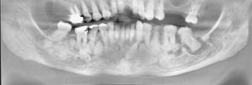
right side (Figure 2). Radiographic images were obtained, and multiple mixed radiolucent-radiopaque lesions
were identified. The central portions of these lesions consisted of sclerotic, hyperostotic
material. These central zones were surrounded by well-defined radiolucencies of various
sizes. The roots of some of the mandibular premolars and molars appeared broadened
and bulbous (Figure 3). Despite these lesions, all of the teeth tested vital with the exception of the
mandibular left first and second molars.

Figure 1Extraoral photograph showing a left-sided facial swelling involving the mandibular
soft tissue and inferior aspect of the nasolabial fold.

Figure 2Intraoral photograph showing expansion of the left mandibular alveolar ridge (A) when compared with the right mandibular alveolar ridge (B).

Figure 3Panoramic radiograph showing multiple mixed radiolucent-radiopaque lesions of the
mandible.
To read this article in full you will need to make a payment
Login with your ADA username and password.
One-time access price info
- For academic or personal research use, select ‘Academic and Personal’
- For corporate R&D use, select ‘Corporate R&D Professionals’
Purchase one-time access:
Reference
Florid cemento-osseous dysplasia: a rare case report evaluated with cone-beam computed tomography.
J Oral Maxillofac Pathol. 2016; 20: 329
Florid cemento-osseous dysplasia: a systematic review.
Dentomaxillofac Radiol. 2003; 32: 141-149
Differentiating early stage florid osseous dysplasia from periapical endodontic lesions: a radiological-based diagnostic algorithm.
BMC Oral Health. 2017; 17: 161
Florid cemento osseous dysplasia in association with dentigerous cyst.
J Oral Maxillofac Pathol. 2010; 14: 63-68
Benign cementoblastoma.
J Oral Maxillofac Pathol. 2011; 15: 358-360
Infected florid osseous dysplasia: clinical and imaging follow-up.
BMJ Case Rep. 2015; 2015bcr2014209099https://doi.org/10.1136/bcr-2014-209099
Clinical, radiographic, and histological findings of florid cemento-osseous dysplasia: a case report.
Imaging Sci Dent. 2011; 41: 139-142
Florid cemento-osseous dysplasia associated with chronic suppurative osteomyelitis and multiple impacted tooth an incidental finding: a rare case report.
J Family Med Prim Care. 2020; 9: 1757-1761https://doi.org/10.4103/jfmpc.jfmpc_1130_19
Florid cemento-osseous dysplasia: a contraindication to orthodontic treatment in compromised areas.
Dental Press J Orthod. 2018; 23: 26-34
Gardner syndrome. In: StatPearls [Internet]. StatPearls Publishing.
Gardner’s syndrome.
J Clin Imaging Sci. 2011; 1: 65
Gardner’s syndrome: a case report and review of the literature.
World J Gastroenterol. 2005; 11: 5408-5411
Adult Paget’s disease of bone.
Clin Med (Lond). 2020; 20: 568-571
Paget disease. In: StatPearls [Internet]. StatPearls Publishing.
Paget’s disease of the mandible.
J Oral Maxillofac Pathol. 2012; 16: 107-109
How to evaluate the activity of Paget’s disease in clinical practice and which patients should be treated? Article in French.
Rev Rhum Mal Osteoartic. 1984; 51: 463-468
Familial gigantiform cementoma: case report of an unusual clinical manifestation and possible mechanism related to “calcium steal disorder.
Medicine (Baltimore). 2016; 95e2956https://doi.org/10.1097/MD.0000000000002956
Management strategy in patient with familial gigantiform cementoma: a case report and analysis of the literature.
Medicine (Baltimore). 2017; 96e9138https://doi.org/10.1097/MD.0000000000009138
A family of familial gigantiform cementoma: clinical study.
J Maxillofac Oral Surg. 2022; 21: 44-50https://doi.org/10.1007/s12663-021-01515-2
Gigantiform cementoma: clinicopathologic presentation of 3 cases.
Oral Surg Oral Med Oral Pathol Oral Radiol Endod. 2001; 91: 438-444https://doi.org/10.1067/moe.2001.113108
Chronic recurrent multifocal osteomyelitis (CRMO): presentation, pathogenesis, and treatment.
Curr Osteoporos Rep. 2017; 15: 542-554
Chronic recurrent multifocal osteomyelitis involving the mandible: case reports and review of the literature.
Dentomaxillofac Radiol. 2010; 39: 184-190
Biography
Mr. Rosen is a student, Columbia University College of Dental Medicine, New York, NY.
Biography
Dr. Sarmiento is a clinical associate professor, Department of Periodontics, University of Pennsylvania School of Dental Medicine, Philadelphia, PA, and in private practice, New York, NY.
Biography
Dr. Rosen is a clinical professor, Department of Periodontics, Rutgers University School of Dental Medicine, Newark, NJ, and in private practice, New York, NY.
Biography
Dr. Peters is an assistant professor, Division of Oral and Maxillofacial Pathology, Columbia University Irving Medical Center, New York, NY.
Article Info
Publication History
Published online: December 19, 2022
Publication stage
In Press Corrected Proof
Footnotes
Disclosures. None of the authors reported any disclosures.
Diagnostic Challenge is published in collaboration with the American Academy of Oral and Maxillofacial Pathology and the American Academy of Oral Medicine.
Identification
DOI: https://doi.org/10.1016/j.adaj.2022.11.002
Copyright
© 2022 American Dental Association. All rights reserved.
ScienceDirect
Access this article on ScienceDirect


