

Drs. Laura Nicolas, Alberto Teramoto, and Manuel Hinojosa discuss how segmentation can provide an essential diagnostic function
Introduction
An adequate treatment plan for resolving impacted teeth could be very difficult if the necessary information is not available. It is mandatory to know the exact location and, more importantly, the immediate relationships of the impacted teeth to surrounding teeth, roots, and even the mandibular nerve.
After the third molars, maxillary canines have the highest frequency of impacted localization, with a prevalence ranging from 1% to 3%,1-4 and are more frequent in female patients by a ratio of 2:1.5 Surgical treatment, when necessary, is difficult and time-consuming when the clinician doesn’t have enough information. Thus, the accurate localization of impacted maxillary canines is very important when the clinician has to decide to treat orthodontically and especially if surgical intervention is required.
Most of the time in daily practice, the first radiographic image required to support the clinical examination is the panoramic radiograph. And sometimes in order to have a better localization of impacted teeth, clinicians can improve diagnosis by using a combination of two or more bidimensional images — occlusal and periapical, which allow the localization of impacted canines, treatment planning, and evaluation of the treatment result.
Importance of CBCT
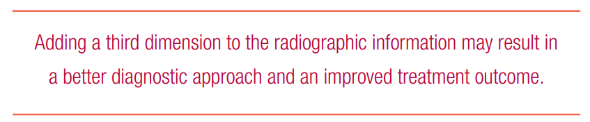
It is well-known that the diagnostic accuracy of these bidimensional radiographic techniques presents many limitations, increasing the risk of mistakes. Adding a third dimension to the radiographic information may result in a better diagnostic approach and an improved treatment outcome — using cone beam computed tomography (CBCT), clinicians can take advantage of 3D information provided by a low radiation dose and at relatively low cost. CBCT provides information that is not revealed during traditional radiographic analysis and is therefore indicated in case of impacted teeth or cranio-facial structural anomalies.
Sometimes even all of the 3D images are not enough because clinicians can use the CBCT data more efficiently. Another use of DICOM data is using segmentation — a process that separates structures of interest from the background and from each other. This process provides an essential analysis function for which numerous algorithms have been developed in the field of image processing; segmentation of an object is achieved either by identifying all pixels or voxels that belong to the object or by locating those that form its boundary. Segmenting teeth from CBCT images is not an easy procedure. Complications include the following:
- DICOM data is acquired with the upper-lower jaw in occlusion, so it is hard to separate a tooth from its opposing teeth along their occlusal surface because of the lack of changes in gray values.
- Separating a tooth from alveolar bone with similar densities is difficult.
- Identifying and differentiating various teeth types is difficult when teeth possess a similar shape.
Some open-source software for segmentation is available on the market; however, the software entails a long learning curve and time to perform a high-quality segmentation.
Recently, Artificial Intelligence technology, Orca Dental AI, created a unique system in a 3D-controllable STL format that offers not only a teeth segmentation service, but also cephalometric and airway volume analyses and videos of teeth segmentation, making this process much more complete, easy to use, and completely informative in disclosing all the impacted relationships.
For a better diagnostic approach and an improved treatment outcome, with the available data, it is also possible to convert the DICOM files of the CBCT to a model using a 3D printer (Zenith D DENTIS CO. LTD., La Palma, California). This very useful tool helps visualize and provide 3D visualization of conditions of impacted teeth and can help the clinician choose the best treatment plan. For example, it can offer more information on whether to perform orthodontic treatment to align an ectopic canine or whether the patient needs a surgical procedure because of the difficulty of aligning ectopic maxillary canines.
Cases
Case 1
Because the position of the impacted canine root is close to the vestibular side, with extraction of the first premolar, it is easy to move the canine to its ideal position (Figures 1A-1H).
Figure 1A (left): Initial intraoral photos and Figure 1B (right): Panoramic X-ray
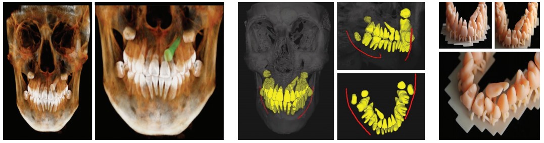
Figures 1C-1E: 1C. Impacted upper canine CBCT image. 1D. Impacted upper canine segmentation (ORCA Dental AI). 1E. Impacted upper canine 3D impression
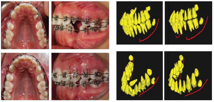
Figures 1F-1H: 1F. Leveling and aligning of impacted canine in 4 months. 1G and 1H. CBCT Segmentation — before and after (ORCA Dental AI)
Case 2
The horizontal-position surgical option is the best choice for this right impacted canine. This is more obvious with this imaging option (Figures 2A-2F).
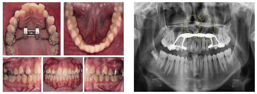
Figure 2A (left): Intraoral photos and Figure 2B (right): Panoramic X-ray
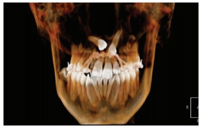
Figure 2C: CBCT image of bilateral impacted canines
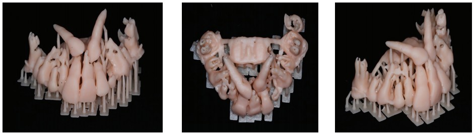
Figures 2D-2F: 2D. Impacted right and left 3D impressions — frontal view. 2E. Impacted right and left 3D impressions — occlusal view. 2F. Impacted right and left 3D impressions — right side view
 Conclusions
 Conclusions
CBCT is a useful tool for clinical situations such as impacted canines, craniofacial anomalies, TMJ assessment, and upper airway analysis. CBCT images in combination with 3D segmentation, impressions, and videos provide a better diagnosis and are useful in deciding whether to treat these cases orthodontically or surgically.
Acknowledgments
Special thanks to Orca Dental AI for the segmentation process in STL format, video, airway, and cephalometric analysis and to Ideas Dentales, Mexico City, Mexico, for printing 3D models.