In this study, we demonstrated that accurate and precise 3D cephalometric analysis is feasible using non-ionizing, high-resolution MRI. In vitro investigations on a phantom showed high concordance between landmark-based MRI measurements and corresponding true values in different phantom positions. In vivo, landmark-based 3D cephalometric measurements as applied in clinical routine revealed high levels of reproducibility, independently from the head position of study participants. Our findings indicate that 3D cephalometric analysis could be performed using MRI in the future. This may have a major impact on planning and monitoring of treatment in orthodontic and orthognathic patients, since MRI scans can be performed repeatedly and independently from the extent of malocclusions without radiation exposure to the predominantly young patients.
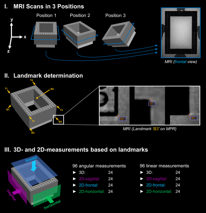
A key finding of this study was that high measurement accuracy could be demonstrated for the applied MRI technique. Independent from the phantom position, all angular and linear measurements revealed a high concordance with the corresponding true values. Precisely, we found mean differences between true values and MRI ranging from −0.12–0.08 mm for 3D distances, −0.22–0.24 mm for 2D distances, 0.00–0.04° for 3D angles and −0.09–0.04° for 2D angles. This is in line with previous phantom studies analyzing the accuracy of 3D MRI methods designed for craniofacial imaging. Goto et al. showed minor differences of 0.2, −0.6 and −0.3 mm between ground truth and 3D MRI measurements using tube phantoms. These differences were only slightly larger compared to identical measurements on CT revealing differences of −0.1, 0.3 and 0.3 mm19. Eley et al. also demonstrated submillimeter discrepancy between true values and 3D MRI at 3 Tesla for 11 linear measurements performed on a cube phantom, with mean values ranging from 0.02–0.73 mm. In the same study, CT was even slightly less accurate, with ranges from 0.03–0.91 mm compared to ground truth22. Overall, our in vitro results are in accordance with these studies and confirm high measurement accuracy of our 3D MRI technique which is crucial for the interpretation of subsequent in vivo measurements.
MRI offers the unique possibility to repeat in vivo examinations for 3D cephalometry as often as required. Given these conditions, we performed 5 MRI examinations in 3 study participants, with different head positions for each scan. Our results revealed a high concordance between repeated measurements in all volunteers. Ranges remained below 2° and 2 mm in all repeated measurements in every study participant. Average ranges were 0.88° for all angles and 0.87 mm for all distances, and if only 3D measurements are included, average ranges were 0.84° and 0.78 mm, respectively. A direct comparison of our results to previous studies is not possible for two reasons: First, comparable MRI studies have not been performed before. Second, for reasons of radiation protection, X-ray based modalities cannot be used to investigate the effect of head positioning on cephalometric measurements in vivo. Consequently, cadaver studies using CBCT to analyze 3D cephalometric measurements at varying head orientations are most suitable for comparison. For instance, Ludlow et al. performed 4 linear cephalometric measurements on 28 dry skulls in 3 positions (ideal, shifted and rotated), and in comparison to direct skull measurements the absolute value of difference ranged between 0.96 mm and 1.94 mm23. Similarly, a CBCT study by Hassan et al. assessed 10 linear cephalometric measurements in two different head positions using 8 dry skulls. Their results showed no statistical difference between the two scan positions, with mean absolute differences between CBCT and direct physical measurements ranging from 0.11–0.39 mm for the ideal scan position and 0.10–0.43 mm for the rotated scan position24. Overall, results of these cadaver CBCT studies correspond very well with the differences between repeated measurements observed in our in vivo MRI study, and thus high in vivo reproducibility can be concluded. Importantly, our data for the first time provides in vivo evidence that head positioning does not have a significant impact on measurement results in landmark-based 3D cephalometry. Under these favorable conditions, MRI not only provides the possibility to perform longitudinal studies for treatment monitoring but also to specifically examine healthy subjects for the establishment of reference values.
Even though associated with several diagnostic limitations of projection radiography1,2,3,4, 2D lateral cephalometric analysis will still be used in clinical routine in the future, as various well-established methods and normative data are available25,26,27,28,29. Previous in vivo studies have already demonstrated that 2D cephalometric analysis can be performed on MRI: In comparison between MRI and lateral cephalometric radiographs (LCR), no clinically relevant discrepancies were observed for 2D analyses including midsagittal16,30 as well as bilateral16 landmarks. Therefore, lateral 2D measurements defined by a 3-landmark-based midsagittal plane (MSP)31 were integrated in the analysis protocol of the present study. This approach revealed a high reliability in repeated 2D measurements with average ranges between minimum and maximum values of 1.17° and 1.08 mm, respectively. From this it can be concluded that high-resolution 3D MR images allow for reproducible lateral cephalometric analysis in vivo. As discussed above, our results also demonstrate that the calculated 3D and 2D values do not depend on head orientation. This is a major advantage of 2D MRI measurements in comparison to 2D measurements on cephalometric radiographs, which are susceptible to measurement errors caused by head rotation2,32,33.
In view of the results of the present study, it is particularly important to discuss how MRI-based 3D cephalometry could be integrated into clinical practice in the future. Until recently, it was believed that MRI cannot serve as a diagnostic modality for planning of orthodontic therapy or orthognathic surgery34. Along with recent technical developments, however, new perspectives have emerged. By combining the latest MRI techniques, we could establish a robust imaging protocol designed for applicability in clinical routine, yielding isotropic images with high resolution and excellent contrast. This was accomplished by using high field MRI (3 Tesla), a 16-channel surface coil and an application-optimized prototype 3D sequence with high spatial resolution. Importantly, the acquisition time of this sequence is only 7:01 minutes and the total examination time lies within approximately 10 minutes including positioning and localizer sequences. Consequently, all in vivo scans could be performed time-efficiently with high comfort and no relevant motion artifacts were observed. The acquired high-resolution 3D images allowed a clear depiction of all predefined cephalometric landmarks, resulting in high in vivo reproducibility. Analysis of MR images was performed by determining cephalometric landmarks on multiplanar reconstructions (MPR), which means the workflow is identical to MPR-based 3D cephalometric analysis on CT or CBCT images. As with CT/CBCT, the time required for landmark determination depends on the number of predefined landmarks as well as the observer’s training and experience. In the present study, in vivo 3D cephalometric analysis included 27 landmarks and was performed by an experienced dentomaxillofacial radiologist within 10–15 minutes per dataset. Altogether, the applied MRI technique enables time-efficient acquisition and analysis of images, thus providing the basis for clinical application of landmark-based 3D cephalometry. As this paper presents a new approach of MRI-based cephalometry with specific technical requirements, availability is limited at this stage. In principle, however, the technique could be established on different MRI systems with similar features for broad clinical use in the future.
Since 3D imaging can substantially improve diagnostic possibilities in orthodontics and orthognathic surgery, many studies have investigated the use of conventional CT and CBCT for 3D measurements of craniofacial structures35,36,37,38,39. Recently, particularly CBCT has moved into focus and proven to be an accurate modality for 3D cephalometric analysis5,6,7. However, the use of CBCT for 3D cephalometry is limited because of considerable radiation doses and a substantially increased lifetime attributable cancer risk of young patients40,41. As a consequence, reference values for 3D cephalometry are not available until today. In contrast to CBCT, MRI is an imaging modality allowing radiation-free 3D imaging of the craniofacial region which could provide a wide range of new diagnostic options. In view of our results and former in vitro studies demonstrating high concordance between measurements on CBCT and MRI20,21, these two modalities might deliver equivalent results for 3D cephalometric analysis in vivo as well (within clinically acceptable margins).
From a methodological point of view, it is important to stress that artifacts caused by metallic materials (e. g. fixed orthodontic appliances, dental implants or osteosynthesis material) can be a limiting factor of MRI in the craniofacial area42. In orthodontics and orthognathic surgery, this could be particularly important for treatment monitoring. To minimize this potential limitation for future patient studies, we used a 3D MSVAT-SPACE sequence which has proven to significantly reduce metal-induced artifacts15.
In conclusion, this study demonstrates that high-resolution MRI based on a short examination protocol can be used for 3D cephalometric analysis. The applied MRI technique revealed an excellent accuracy in vitro and high levels of reproducibility in vivo, independently from the position of investigated object/head. Thus, non-ionizing MRI has the potential to overcome the limitations of X-ray based standard methods, which are the limited diagnostic value of conventional radiographs and the radiation risks associated with CT and CBCT. In absence of radiation exposure, MRI offers the possibility to repeatedly examine patients with varying degrees of orthodontic disorders as well as healthy subjects, which might substantially contribute to provide evidence for the diagnostic and therapeutic efficacy of 3D cephalometry.


