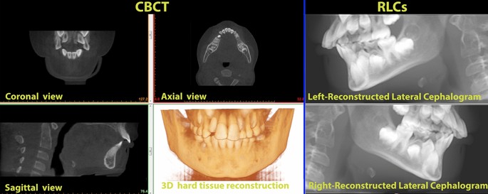
The analysis of mandibular growth changes during the pubertal growth spurt has important consequences for the diagnosis and treatment planning of skeletal malocclusions. The majority of knowledge regarding facial growth comes from bi-dimensional cephalometric tracings and their superimposition25,26,27. Evaluation of growth and the changes caused by growth using bi-dimensional radiographs raise an important issue as a 3D object is flattened to a bi-dimensional image14.
The first aim of this study was to test whether mandibular growth in skeletal class 2 patients assessed on CBCT and on traditional 2D cephalometry showed any difference. The second was to test whether the error in bi-dimensional cephalograms was linked to the ratio between transverse and sagittal growth of the mandible. The results of the present study regarding average mandibular growth (Table 1) are in concordance with those published by Gomes and Lima28 that reported an annual mandibular body growth rate of as 2.16 mm/year during the pubertal growth spurt of skeletal class 2 patients.
The decision to select a sample of skeletal class 2 patients is because it appears to be the most frequently occurring malocclusion, especially in North America and Europe29,30,31. During orthodontic treatment, clinicians need to monitor mandibular growth, and this is enabled by longitudinal evaluation of lateral teleradiographs.
However, bi-dimensional measurements of craniofacial structures that do not lie on the mid-sagittal plane present various degrees of distortion, and the evaluation of growth could be affected. As suggested by Farronato et al.32 the comparison of the tracings from bi-dimensional radiographs and 3D CBCT scans shows that the measurement of the segment Go–Me (representing mandibular body) presents a statistically significant difference. Pittayapat et al.33 reported that cephalometric values measured on bi-dimensional radiographs are expected to be significantly different compared to those measured on CBCT. The segment Go–Me, representing mandibular body length, is the most affected by projection distortion33. This result can be explained by the fact that 2D radiographs are obtained by flattening a 3D object, thus projecting and distorting each point in proportion to its distance from the mid-sagittal plane. The mandibular body lies angled to the mid-sagittal plane, thus resulting in strong underestimation when projected on a bi-dimensional plane.
In this study, at each point in time, linear measurements of mandibular length were found to be significantly lower in RLCs than in CBCT by approximately 12 mm on both sides (Table 2).
To better understand the reason behind our choice to investigate this specific topic, one must consider the physiological pattern of mandibular growth. Mandibular growth has been extensively studied by researchers such as Enlow11,12, Franchi26,34 and Bjiork3,9,10. Significant morphological changes have been reported in the mandible during the peak of growth35. The available literature on the subject suggests that the mandible has different growth sites during growth spurt3,10,11,12,26. The main growth site is at the level of the mandibular condyle3,10,11,12,26.
The surface of the condyle grows with endochondral ossification, resulting in an increase mainly in the vertical dimension. Nonetheless, other sites are active during this period. For example, bone remodelling at the level of the mandibular body takes place at the level of the gonial angle; more precisely, the translation of the mandible forward and the increase in the sagittal dimension are due to the apposition of bone on the posterior surface of the mandibular ramus3,9,10,11,12. The mandibular body lengthens through the process of periosteal ossification with bone resorption on the anterior side of the mandibular ramus and contemporary bone apposition on the posterior surface of the mandibular ramus3,9,10,11,12. During the first years of life, the anterior surface of the ramus is located around the point where the first milk molar erupts5,36. The progressive bone resorption and apposition at the level of the ramus determines the growth of the body and the space available to accommodate the permanent molars. In the case of posterior dentoalveolar discrepancy and retention of mandibular molars, one possible explanation is indeed a reduced resorption at the level of the ramus37. As the growth of the mandibular body occurs posteriorly with divergent progression, it also involves an increase in the transverse dimension.
As mandibular growth appears to be a tri-dimensional phenomenon, the most accurate examination to assess mandibular body changes during adolescence is superimposing tri-dimensional volumes of the mandible obtained with a CBCT scan by tri-dimensional image registration based on the correspondence of voxels34,38. In fact, several authors are trying to identify stable points at the mandibular level to overlap the jaws and evaluate growth in a reliable way34,39. However, the justification and optimization principles that rule radiation exposure strongly limit the use of CBCT and therefore the available data as reported by the SEDENTEX and DIMITRA guidelines16,40,41. The potential benefits of this type of examination should be weighed against the increased exposure to ionizing radiation compared to conventional bi-dimensional imaging. Avoidance of unnecessary radiation exposure is crucial, particularly for children and adolescents40,41.
Contrary to the authors’ expectations, mandibular growth did not show any significant difference between RLCs and CBCT as shown in Table 2. This event seems to be attributable to the pattern of mandible growth. As reported in Table 1, the transverse growth of the mandible (ΔGoR–GoL) is similar to sagittal growth (ΔGo–Me), as indicated by the lack of a statistically significant difference between the angles GoR–Mê–GoL in the two points in time (see Table 1), thus showing a similar distortion in both RCLs. To further investigate the concordance of the measurements of mandibular growth in the two scans, a correlation analysis was carried out (see Table 5). Linear regression between mandibular growth in 3D and 2D (the independent and dependent variables, respectively) showed a very high correlation (R2 = 0.969), a beta coefficient close to 1 (Δ_3D coefficient = 0.984; p < 0.001) and a non-statistically significant constant (constant = 0.032 mm; p = 0.848). These values testify to how accurately RLCs are compared to CBCT and how little the values differ.
When growth is evaluated longitudinally, the least noticeable difference defines the threshold for growth detection. Our results show that measurements of the mandibular body length and its growth with CBCT are comparable to those of RLCs in terms of precision (see Table 3). Cephalometric measurements taken on RLCs were highly reliable, as inter-observer and intra-observer agreement was excellent and slightly lower than those measured on CBCT.
The Bland–Altman test for mandibular length showed that 95% limits of agreement of both imaging methods (CBCT, RLC) were from 15.17 to 15.97 mm for the upper bound and from 7.43 to 8.50 mm for the lower bound. Furthermore, mandibular body lengths assessed on CBCT were systemically larger than those measured on RLC. The average mean differences between CBCT and RLCs at T0 were 11.54 mm (right side) and 11.70 mm (left side); at T1, the average mean differences were 11.91 mm (right side) and 11.89 mm (left side). This difference is due to the bi-dimensional flattening of the skull in RLCs and its aforementioned consequences.
The Bland–Altman test for mandibular growth (Fig. 2 and Table 4) showed that the 95% limits of agreement of both imaging methods (CBCT, RLC) were instead − 1.14 mm (right side), − 1.31 mm (left side) and − 0.78 mm (on average) for the lower bound and 1.19 mm (right side), 0.57 mm (left side) and − 0.448 mm (on average) for the upper bound. The measurements of mandibular growth made with CBCT and RLC were very close: the mean differences were 0.03 (right side), − 0.37 mm (left side) and − 0.17 mm (average).
Since the measurements were performed by two operators, the evaluation of all four measurements (two measurements from each observer) in the Bland–Altman plot analysis considered the entire variation, thus making the limits of agreement wider and more realistic.
Considering the overall high concordance with CBCT and the low radiation exposure, lateral cephalometric analysis for the assessment and monitoring of mandibular growth can be performed by lateral cephalograms without any difference from the radio-diagnostic point of view.
Some authors are attempting to validate an MRI-based cephalometric protocol, which could have an important effect on treatment planning and the possibility of monitoring mandibular growth longitudinally, especially in the case of young patients, since MRI can be repeated due to the absence of ionizing radiation42,43,44,45. The current development and spread of radiation-free technologies in dentistry, such as MRI, will allow researchers to better understand maxillo-facial growth and physiology46,47,48.
Focusing on the other aim of this study, which is whether mandibular reshaping during pubertal growth spurt could interfere with mandibular growth assessment through bi-dimensional radiographs, the second linear regression has a twofold meaning. There is a meaningful relationship (R2 = 0.81) between the bi-dimensional error in assessing growth and the mandibular growth pattern (the ratio or transverse to sagittal growth). If the transverse component of growth is greater than the sagittal component, the resulting distortion in the second radiograph will lead to an underestimation of the real growth, as the projection of the mandibular body is affected by a greater OAB angle (Fig. 4). In contrast, if the reverse is true and the sagittal component is greater, the bi-dimensional growth will appear larger than the real growth assessed on CBCT. However, the OAB angle, which is half of the mandibular symphyseal angle (assuming these patients’ mandibles are almost perfectly symmetrical), does not show any statistically significant difference between the two points in time even though it appears that in the majority of patients, it trends toward a slight and not clinically significant reduction (− 0.86 ± 2.41). In fact, the second linear regression (coefficient of ratio = − 1.850, p < 0.001; constant = 0.517, p < 0.001) shows that in the cohort of patients who was analysed, the expected value of distortion in the assessment of mandibular growth ranges between − 0.732 and 0.265 mm, thus showing a result that is not clinically significant.
(a) Cephalometric landmarks used in the present study. For each CBCT and RLC dataset, 3 cephalometric landmarks were determined on multiplanar images. Definitions of cephalometric landmarks: GoL/GoR = left/right gonion (midpoint on the curvature of the angle of the mandible); Me = menton (most inferior point of mandibular symphysis). Definitions of cephalometric measurements: GoL/GoR-Me = mandibular body length right/left; GoL-GoR = inter-gonial distance; GoR–Mê–GoL = mandibular symphyseal angle. (b) Isosceles triangle (CAB) representing the mandible of the patient. The authors considered an isosceles triangle for each patient having the symmetrical sides (AB; AC). The isosceles triangles were divided in two equal right triangles (ABO). Definitions of the measurements: AB/AC (hypotenuse) = right/left mandibular body length; BC = inter-gonial distance; BO (cathetus) = half inter-gonial distance and transverse component of the mandible; AO (cathetus) = sagittal component of the mandible; CÂB = mandibular symphyseal angle; OÂB = half mandibular symphyseal angle.
Lin et al.49 affirmed that significant errors exist in the measurements of mandibular body growth using 2D RLCs compared to CBCT in six miniature pigs. Clinical and statistically significant underestimation of the actual growth was reported. Lin et al.49 concluded that this event should be considered in orthodontic treatment planning to more precisely control the treatment process and outcome. However, the present study reveals that within its limitations, the actual linear mandibular body growth in humans during growth spurt is equally evaluated in entity and accuracy with bi-dimensional radiographs. These phenomena occur because humans and pigs have different mandibular development, and no generalization should be made based on their data. The development of the human mandible appears to be completely different, as it develops almost by the same amount in the sagittal and transverse planes, keeping the distortion between 2 and 3D stable at different points in time. It is therefore obvious that although the second regression analysis points to a close relation between growth pattern and projection distortion, the amount of error is of no relevance from any point of view. Bi-dimensional assessment of linear mandibular growth should therefore be performed accurately on lateral cephalograms, which are the absolute gold standard and have been for almost a century.
Conventional 2D imaging methods are inherently limited by anatomical superimpositions and inherent image distortion14. However, the authors confidently affirm, within the limits of the current study, that mandibular growth shows no clinical or statistical difference whether it is assessed on conventional lateral cephalograms or on CBCT.
Measured values appear equally reliable and precise. Although a difference exists, the difference in mandibular growth as assessed with the two imaging methods is of no clinical or statistical significance. The development of the human mandibular body through puberty appears to be almost equal in the transverse and sagittal planes, thus keeping distortion of the mandibular body similar between CBCT and conventional bi-dimensional radiographs at the two analysed points of time.
One of the weaknesses of the study is its sample size, which is small, albeit adequate to provide a sufficient level of confidence in the results in accordance to the sample size calculation (see dedicate section: “Statistical analysis and sample size calculation”). The present study investigates only the changes occurring during the peak of growth; further investigations should focus on morphological changes occurring before and after it. Another possible development of the present study could include the analysis of mandibular growth represented as the distance between the Condylion point (representing the highest and most posterior point on the contour of the mandibular condyle) and menton. However, the study design would be much more complex and could be affected by many biases, as it would be difficult to construct a simplified model that considers the influence of both the change in the angle of the menton and the change in the angle between the mandibular body and mandibular ramus. The analysis of the effect of facial orthopaedic treatment of mandibular shape could also prove to be of some interest. The evaluation of patients with different skeletal classes and those presenting various degrees of asymmetry would be beneficial. Such data would allow us to draw conclusions that could more easily be extended to the entire population, as the findings of the present study fit only people who present a satisfactory degree of facial symmetry.


