Subjects
The subject included 98 patients who searched for orthodontic or orthognathic treatment in the hospital. Among the patients, 28 were male and 70 were female. The age ranges from 8 to 42 with an average age 23.63 ± 6.64. The criteria for the inclusion of patients were:
- 1)
No habits of smoking and/or drinking;
- 2)
No exposure to X-rays in recent three months;
- 3)
No oral mucosa diseases;
- 4)
No local stimulation factors;
- 5)
would have dental X-ray examinations within 1 hour.
When not all the above inclusion criteria are met, a patient was excluded.
Altogether, 24 patients for orthognathic surgical treatment and 74 patients for orthodontic treatment were collected. Prior to X-ray examinations, patient individual information such as age, gender, medical history, radiographs exposed and the exposure parameters were recorded for later analysis. All the acquired radiographs were those necessary for treatment planning and not specific for the study.
An information consent form was signed by the participants or their guardians. The study was approved by the Institutional Review Board of Peking University School and Hospital of Stomatology. All methods were performed in accordance with the relevant guidelines and regulations.
Dental X-ray examinations
One or several of the following dental X-rays were performed for individual patient: panoramic radiograph, lateral cephalometric radiograph, posteroanterior cephalometric radiograph, a CBCT scan for TMJ, a CBCT scan for the whole skull and a CBCT scan for maxilla. CBCT scans were performed with a DCT Pro scanner (VATECH&E-WOO Corporation, Seoul, Korea; 90 kVp, 5–7 mA, 24 s) or a NewTom VG (Quantitative Radiology, Verona, Italy; 110 kVp, 6.24–14.45 mAs). Panoramic radiograph, lateral radiograph and posteroanterior radiograph were performed with an all-in-one dental digital imaging system Orthopantomograph OP100 (Instrumentarium Imaging Corporation, Tuusula, Finland). The exposure parameters were 66 kVp, 4–10 mA, 17.6 s for the panoramic radiographs and 77 kVp, 12 mA, 0.5–1.0 s for the lateral radiographs, 77 kVp, 12 mA, 0.8–1.2 s for the posteroanterior radiographs.
Cell collection and slides preparation
Exfoliated oral mucosa cells were collected immediately before dental X-ray examinations and 10 days later. After rinsing the mouth with tap water, cells were obtained by swabbing both left and right cheek mucosa of the patients with a moistened wooden spatula. Cells were transferred to a tube of buccal cell buffer (0.16% Tris-HCL, 0.12% EDTA, 3.72% sodium chloride), centrifuged three times (2000 rpm, 3 min), fixed in 3:1 methanol/acetic acid, homogenized for 5 min, dropped onto pre-cleaned slides and dried in air. Slides were successively stained with the method of Feulgen/fast green.
Cytological observation
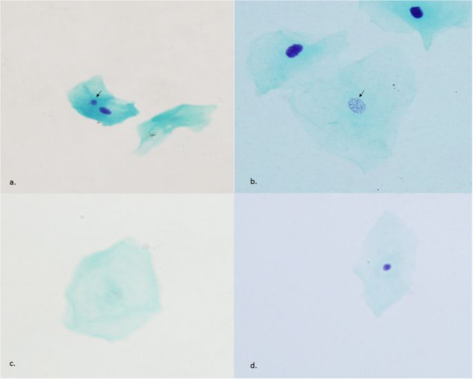
Cytological observation was performed with a light microscope BX51 (Olympus corporation, Tokyo, Japan) at x400 magnification. The frequency of micronuclei cells was counted in 2000 cells and other types of cells such as basal cells, binucleated cells, condensed chromatin cells, karyorrhectic cells, pyknotic cells, karyolytic cells and cells with nuclear buds were scored in 1000 cells for each individual. All kinds of cells were scored according to the criteria described by Thomas P et al.4. Micronuclei cells as a parameter for DNA damage were distinguished on the basis of five characteristics: (a) be less than 1/3 diameter of the main nucleus; (b) be on the same plane of focus; (c) have the same color, texture and refraction as the main nucleus; (d) have smooth round or oval shape, and (e) be clearly separated from the main nucleus9. Sample images of the observed MN and other cells were shown in Fig. 1.
Photomicrographs of cells with (a) micronucleus (arrow), (b) karyorrhexis (arrow), (c) karyolysis and (d) pyknosis. Magnification x400.
For intra-observer variability analysis, ten slides were randomly selected for observation of cell anomalies two month later.
To study whether the rate changes of different types of cells are really connected to dental x ray examinations, 8 patients were called back one and half year later and had the mucosa cells collected for cytogenetic observation again.
Measurement of accumulated absorbed doses of oral mucosa
An human anthropomorphic phantom (ART-210, Radiology Support Device, Inc., Long Beach, CA, USA.) and thermoluminescent dosimeter (TLD) chips were used for the estimation of accumulated absorbed dose of oral mucosa. The phantom was with tissue equivalent X-ray attenuating characteristics and closely conforms to International Commission on Radiation Units and Measurements specifications10.
The accumulated absorbed doses of oral mucosa were measured at each of the nine protocols described in Table 1. Before the study, all dosimeters were calibrated using a Co-60 source. Detailed information regarding the measurements was described in a previous study11.
Statistical analysis
Software package SPSS v16.0 for windows (SPSS, Chicago, IL, USA) was employed for the statistical analysis. Differences between frequencies of micronuclei cells, basal cells, binucleated cells, condensed chromatin cells, karyorrhectic cells, Pyknotic cells, Karyolytic cells and cells with nuclear buds before and after X-ray examinations were analyzed by Wilcoxon signed rank test. Since the basic images required for an orthodontic or orthognathic surgical treatment are 2 dimensional planar images, the absorbed doses were accordingly divided into two groups, low and large dose groups, for further analysis. Wilcoxon signed rank test was also used to analyze different cell rates before and after X-ray examinations in both the large and the low dose groups. For the analysis of correlation among different absorbed dose levels and changes of cell rates, Spearman rank correlation was employed. The changes of cell rates is the difference in the counted cell rates before and after radiographic examination (change of cell rates = Cell rate after X-ray examinations – Cell rate prior to X-ray examinations). According to age 18, the patients was divided into two groups, that is, in one group the patients was younger than 18 years old and in the other group the patients were older or equal to 18 years old. To investigate the age effect on the observed cell anomalies, the change rates obtained from both the low and large dose group were analyzed by Mann-Whitney test. For intra-observer variability, one-way ANOVA was employed. Differences were considered to be statistically significant when P < 0.05.


