The effect of ethnicities on the root and canal morphology of mandibular second molar is well documented in the literature4, 5, 15. Furthermore, mandibular second molar is the most common tooth to exhibit C-shaped canal morphology3. Here we investigated the root and canal morphology of mandibular second molar in Emirati population. We have also described and studied the changes in the C-shape configuration along the root length. This is an effort to address the knowledge gap related to this population and an attempt to describe the C-shaped molars as a single unit.
In the present study the most common root morphology in mandibular second molars was two separate roots (78.3%), one mesial and one distal. The other types of root morphology observed were molars with single conical root (2.4%), three roots (0.8%) and four roots (0.6%). Similar findings of presence of two roots followed by 3 roots in mandibular second molars have been reported in several studies16, 17. The prevalence of three rooted mandibular second molar documented in other populations ranges from 0.26 to 8.98%5, 18, 19. The third root observed in our study was found lingually in 75% of cases, which is in agreement with reports in other population17, 20, 21. Therefore, despite the low prevalence, the clinicians should be aware of such morphological variations in the number of roots, to successfully manage endodontic treatment in these cases.
According to our findings, Type I VC (90.5%) was the most prevalent configuration in the distal root. However, Type II (46.5%) and III (39.4%) VC were the most common root canal configurations in the mesial root. In contrast to our findings, Type II and Type IV VC were more prevalent in the mesial root of most other studied populations16, 18, 22, 23. The high prevalence of type III VC in mesial root has been reported in very few studies (range 26.2 to 48.45%)15, 24, 25. According to our findings, Vertucci Type VI, VII and VIII were not detected in the sample of mandibular second molars examined. This is in agreement with several other published studies18, 19. Therefore, our results showed that Type II VC is the most common canal configuration in the mesial root in Emirati population. Thus, the clinicians treating this population should pay special attention in managing the mesial root of non-C-shaped mandibular second molar. Modifications of cleaning and shaping technique, maybe considered to prevent tearing the common apical foramen and avoid stripping, ledging and instrument fracture26. Moreover, the second most common canal configuration in the mesial root is Type III VC. Therefore, clinicians should explore for a second canal even if there is one orifice coronally. The inability to detect and treat the second canal might lead to endodontic failure.
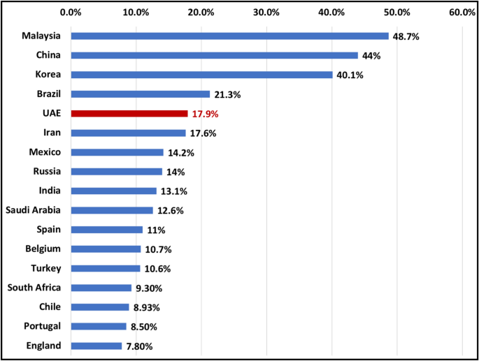
There is a wide variation in prevalence of C-shaped mandibular second molars based on ethnicity and population studied. The prevalence in different populations ranged from 3 to 48.7%5, 19 (Fig. 6). According to our findings, the prevalence of C-shaped mandibular second molars in Emirati population was 17.9%. The prevalence is relatively higher compared to other middle eastern countries such as Saudi Arabia 7.9–12.57%27, 28 and Turkey 4.1–10.6%22 but lower than that of Chinese, Korean and Malaysian population (44–48.7%)5, 19. Moreover, our analysis showed that C-shaped molars were significantly higher in females compared to males (P < 0.005). Our results are in agreement with other studies which reported an association between gender and high prevalence of C-shaped molars4, 5.

Bar chart indicating C-shaped mandibular second molar prevalence in different populations. These studies were conducted using CBCT.
Furthermore, the effect of ethnicity has also been reported on C-shaped canal configuration at different thirds of the root. In Iranian population, the most common canal configuration at the coronal level was C1 (50%), in the middle C3d (32.9%) and apical C3d (36.6%)29. Whereas, in Saudi Arabia, C3c was most common (37.1%) at coronal third, C3c (32.3%) in the middle third and C3d (30.6%) in apical third30. In the present study, the most common canal configuration in C-shaped mandibular molars at the coronal, middle and apical level was C1(41.75%), C3c (51.7%) and C3d (65.9%) respectively.
In the present study, the majority of the C-shaped mandibular second molars had a lingual radicular groove (88%) while only 12% had buccal radicular groove. Our findings are similar to those findings of Jin et al. and Kim et al. in Korean population31, 32, Martin et al. in Portuguese population33 and Alfawaz et al. in Saudi population30, in which the prevalence of buccal radicular groove was less than that of lingual radicular groove (1–22.6% prevalence of buccal radicular groove). In contrast to our findings, Ladeira et al. observed the presence of buccal radicular groove in more than two thirds (69.4%) of the C-shaped mandibular second molars in Brazilian population34. As documented, the dentin thickness is least at the groove area35. Therefore, knowing the location and direction of the groove is important to avoid overpreparation of the canal at that region which can result in iatrogenic perforation.
Moreover, we observed a change in the C-shaped canal configuration coronal to apical in 94.5% of the studied C-shaped mandibular molars. This is similar to the findings of Zheng et al. in Chinese population6. Whereas, the change in canal configuration was observed only in 66% of C-shaped molars in Korean population32. However, the types of morphological change along the length of the root of C-shaped canal configuration was not studied in those populations. Describing the changes in C-shaped canal morphology along the length of the root, can allow researchers and clinicians to have a better understanding of C-Shaped molars as a single unit. This will allow developing new treatment strategies to manage such teeth. Here, we attempted to study such a change and our analysis revealed 4 common types of morphological change in the C-shaped mandibular second molars. Specifically, the most common type was T1: C1-C2-C3d (18%) followed by T2: C1-C3c-C3d (15.4%), T3: C4-C3c-C3d (7.7%) and T4: C3c-C3c-C3d (7.7%). This finding could indicate an effect of ethnicity on presence of specific types of morphological change in C-shaped canal configuration in certain population. However, further studies in other populations are required to confirm such association. Such information if available, can help the clinicians to manage these cases, in addition to other available tools and techniques.
C-shaped canal configuration with the presence of narrow ribbon-like and fan-shaped areas, transverse anastomoses, lateral canals and apical delta make the cleaning and shaping of these teeth challenging2. With the relatively high prevalence in Emirati population, clinicians should consider using advanced tools to diagnose and manage such complex anatomy such as CBCT10, Dental operating microscope36, advanced irrigation activation and delivery systems (such as passive ultrasonic irrigation and negative pressure, laser activated irrigation)37,38,39 and calcium hydroxide as intra canal medicament3. Furthermore, as the most common types of morphological change in C-shaped molar ends up with C3d apically, clinicians should make sure to locate and clean both canals to avoid any failures.
One limitation of our study is that it’s a retrospective study, therefore the inability to control certain factors like FOV, voxel size and the quality of CBCT scan image. Therefore, in the present study, overall image resolution and quality was influenced due to the medium size FOV (8 cm × 8 cm) CBCT scans. However, the voxel size used was 0.15 which is considered acceptable when compared to other studies40. Furthermore, further studies are required in different population to determine the effect of ethnicity on the pattern of change in C-shaped molar along the root length. Another limitation of this retrospective study is that ethnicity was determined based on holding UAE citizenship. Therefore, the data may not represent the whole UAE population, as the UAE nationals represent almost 11% of the total population41,42,43. However, the results of this study are important for clinicians treating UAE nationals, as there is an overall agreement that there is low variation in the ethnic groups among the UAE nationals41.

